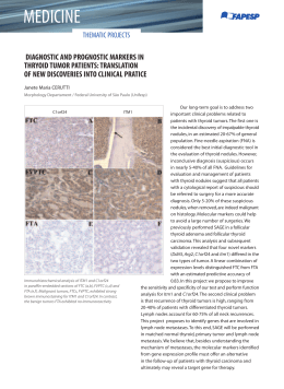
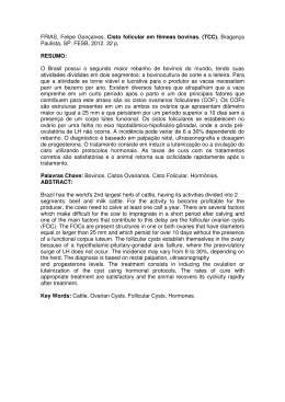
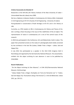
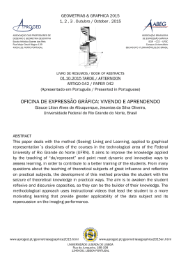
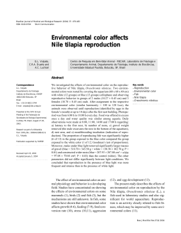
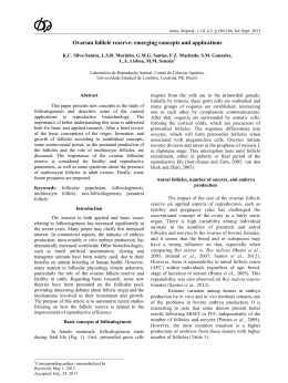
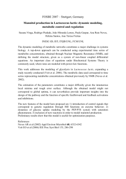
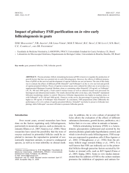
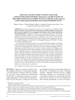
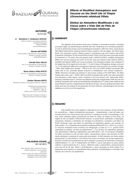
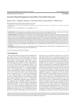
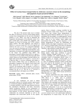
Pesq. Vet. Bras. 35(4):371-376, abril 2015
Morphological and immunohistochemical characterization
of angiogenic and apoptotic factors and the expression of
thyroid receptors in the ovary of tilapia Oreochromis
niloticus in captivity1
Fernanda C. Santos2, Juneo F. Silva2, Jankerle N. Boeloni2, Edgar Teixeira3, Eduardo
M. Turra2, Rogéria Serakides2 and Natália M. Ocarino2*
ABSTRACT.- Santos F.C., Silva J.F., Boeloni J.N., Teixeira E., Turra E.M., Serakides R. & Ocarino N.M. 2015. Morphological and immunohistochemical characterization of angiogenic and apoptotic factors and the expression of thyroid receptors in the ovary of
tilapia Oreochromis niloticus in captivity. Pesquisa Veterinária Brasileira 35(4):371-376.
Escola de Veterinária, Universidade Federal de Minas Gerais, Av. Pres. Antônio Carlos 6627,
Belo Horizonte, MG 31270-901, Brazil. E-mail: [email protected]
Morphological and immunohistochemical characterization of angiogenic and apoptotic
factors and the expression of thyroid receptors in the ovary of tilapia Oreochromis niloticus
in captivity were studied. The morphological evaluation of the ovaries was performed by
histological paraffin embedded and stained with HE. The immunohistochemical expressions of CDC47, VEGF, Flk-1, angiopoietin, Tie-2 and thyroid receptor (TRα) were performed by the technique of streptavidein-biotin-peroxidase. Apoptosis was assessed using
the TUNEL kit. The relative expression of thyroid hormone receptors (TRα and TRβ) was
assessed by RT-PCR real time. The nuclear expression of CDC47 increased with the stage of
maturation of the oocyte and was observed in the follicle cells. Apoptotic bodies were observed in the follicular cells of atretic follicles and postovulatory follicles from the ovaries
of 150g and 350g fish. Expression of VEGF and its receptor Flk-1 was also observed in the
follicular cells, and the expression of both increased with the maturity of the oocyte, with
a higher intensity observed in the full-grown follicle. The expression of angiopoietin and
of its receptor (Tie 2) was discrete and moderate respectively. TRα expression was independent of follicular development. However, the 350 g tilapia exhibited higher expression
of TRβ compared with the 50 g tilapia. We conclude that the proliferative activity and the
expression of VEGF and its receptor increase with follicular maturation and that the TRs
expression increases with ovarian maturity in tilapia (Oreochromis niloticus).
INDEX TERMS: Angiogenic factor, apoptotic factor, thyroid receptors, ovary, tilapia, Oreochromis niloticus, fish, immunohistochemistry.
RESUMO.- [Caracterização morfológica e imuno-histoquímica de fatores angiogênicos e apoptóticos e da expressão de receptores tireoidianos no ovário de tilápia
Received on July 28, 2014.
Accepted for publication on January 25, 2015.
2
Laboratório de Patologia, Departamento de Clínica e Cirurgia Veterinárias, Escola de Veterinária (EV), Universidade Federal de Minas Gerais
(UFMG), Av. Pres. Antônio Carlos 6627, Belo Horizonte, MG 31270-901,
Brazil. E-mails: [email protected], [email protected].
br, [email protected], [email protected], serakidesufmg@
gmail.com; *Corresponding author: [email protected]
3
Laboratório de Aquacultura, EV-UFMG, Av. Pres. Antônio Carlos 6627,
Belo Horizonte, MG 31270-901. E-mail: [email protected]
1
371
Oreochromis niloticus em cativeiro.] Foram estudadas as
caracterizações morfológica e imuno-histoquímica de fatores angiogênicos e apoptóticos e a expressão de receptores
tireoidianos no ovário de tilápia Oreochromis niloticus de
cativeiro. A avaliação morfológica dos ovários foi realizada
por cortes histológicos incluídos em parafina e corados por
HE. As expressões imuno-histoquímicas de CDC47, VEGF
e seu receptor Flk-1, angiopoetina e seu receptor Tie-2 e
recertor tireoidiano (TRα) foram realizadas pela técnica
de estreptavideina-biotina-peroxidade. A apoptose foi avaliada utilizando-se kit de TUNEL. A expressão relativa dos
receptores de hormônios tireoidianos (TRα e TRβ) foi ava-
372
Fernanda C. Santos et al.
liada pela técnica de RT-PCR tempo real. A expressão nuclear de CDC47 aumentou com a fase de maturação do oócito
e foi observada nas células foliculares. Corpos apoptóticos
foram observados nas células foliculares de folículos atrésicos e folículos pós-ovulatórios de ovários de peixes com
150g e 350g. A expressão de VEGF e do seu receptor Flk-1
foi também observada nas células foliculares , e a expressão
de ambos aumentou com a maturidade do oócito , com uma
maior intensidade no folículo maduro. A expressão de angiopoietina e do seu receptor (Tie 2) foi discreta e moderada, respectivamente. A expressão de TRα foi independente
do desenvolvimento folicular. No entanto, a tilápia de 350g
apresentou maior expressão de TRβ em comparação com
a tilápia de 50g. Conclui-se que a atividade proliferativa e
a expressão de VEGF e de seu receptor aumenta com a maturação folicular e que a expressão dos TRs aumenta com
a maturidade do ovário em tilápia (Oreochromis niloticus).
TERMOS DE INDEXAÇÃO: Fator angiogênico, fator apoptótico, receptores tireoidianos, ovário, tilápia, Oreochromis niloticus, peixe,
imuno-histoquímica.
INTRODUCTION
The practice of aquaculture has been growing every year,
and there is an increasing desire to introduce native fish
species in the practice of commercial production. However,
several species of migratory fish, which have high commercial value, do not reproduce spontaneously in captivity
(Santos et. al. 2008a), which has limited the cultivation of
these fish species.
Tilapia (Oreochromis sp.) is a highly prolific species, but
little is known about its ovarian morphophysiology. The
morphology of the female reproductive system is highly variable in teleosts (Tyler 1996). Knowledge of the morphological and physiological characteristics of the ovaries is important for understanding the reproduction of any type of
fish. Thus, understanding the reproductive characteristics
of tilapia, including the proliferative activity, angiogenesis,
apoptosis and expression of thyroid hormones receptors
during oocyte development and during ovary maturation,
may be important for the development of reproductive techniques that permit the maturation and induction of spawning effectively in the migratory species in captive.
The objective of this study was to characterise morphologically, the ovaries of tilapia with respect to proliferative
activity, angiogenesis, apoptosis and expression of thyroid
hormone receptors.
MATERIALS AND METHODS
Eighteen tilapia (Oreochromis niloticus) with weights of 50g, 150g
and 350g grown in captivity and kept in a closed system of recirculating water in the Aquaculture Laboratory (LaQua) from the
Veterinary School of Universidade Federal de Minas Gerais (UFMG)
were used. The system allowed for the on-going maintenance of
water quality and temperature at 26±0.5°C. The variables of water quality, dissolved oxygen, pH and temperature were monitored
daily, and nitrite and total ammonia levels were measured weekly.
The fish were fed a commercial diet with 36% crude protein twice
daily in an amount close to 3% of the stored biomass.
The fish were divided into three groups by weight with six
Pesq. Vet. Bras. 35(4):371-376, abril 2015
animals per group: group 1 consisted of 50 g fish, group 2 consisted of 150g fish, and group 3 consisted of 350g fish. The weights
were chosen to provide the assessment of oocyte development in
several stages. The animals were euthanised according to the protocol approved by the ethics committee on animal experimentation (CETEA/UFMG). Fragments of the right and left ovaries were
fixed in Bouin’s and paraformaldehyde, processed by routine techniques, paraffin embedded and stained with hematoxylin and
eosin for morphological analysis, immunohistochemistry and TUNEL, respectively. Morphologic analysis and the maturation stage
of the oocytes were determined according to the classification recommended by Bazzoli et. al. (2014). Fragments of the right and
left ovaries were also frozen at -80 ºC to determine the relative
expression of thyroid hormone receptors (TRα and TRβ) by real-time RT-PCR.
The ovarian follicles were classified according to the stage of
development as early and late primary growth, early secondary
growth and full-grown follicles, postovulatory follicle and atretic follicles. Morphological analysis was performed descriptively
with respect to the classification of the stage of follicular.
Histological sections of the ovaries were subjected to immunohistochemical analysis to evaluate the proliferative and angiogenic activities and expression of thyroid hormone receptor (TRα)
using the following antibodies: anti-VEGF (vascular endothelial
growth factor: sc-152, Santa Cruz Biotechnology, CA, USA), anti-Flk1 (receptor for vascular endothelial growth factor: sc-6251,
Santa Cruz Biotechnology, CA, USA), anti-angiopoietin 2 (Santa
Cruz Biotechnology, CA, USA), anti-Tie2 (angiopoietin receptor:
Santa Cruz Biotechnology, CA, USA), anti-CDC47 (47DC141, Neomarkers, Fremont, CA, USA), and anti-TRα (α thyroid hormone
receptor: Abcam, Cambridge, UK).
Biotin-streptavidin peroxidase (Streptavidin Peroxidase,
Lab Vision Corp., Fremont, CA, USA) and antigenic recovery
techniques with a retrieval solution were employed for 20 minutes. Histological sections were incubated overnight in a humid
chamber with primary antibodies and were incubated during the
steps of blocking endogenous peroxidase, blocking serum (Ultra
Vision Block, Lab Vision Corp., Fremont, CA, USA) and streptavidin peroxidase for 30 minutes. The primary antibodies and their
dilutions included anti-VEGF (1:400), anti-Flk1 (1:700), anti-angiopoietin 2 (1:30), anti-Tie-2 (1:100), anti-CDC47 (1:100), and
anti-TRα (1:400). Incubation with the secondary antibody (goat
biotin, Lab Vision Corp., Fremont, CA. USA) was performed for
45 minutes. The chromogen utilised was diaminobenzidine (DAB
substrate system, Lab Vision Corp., Fremont, CA. USA). Sections
were counterstained with Harris hematoxylin. The antibodies
used were raised in rabbits. To check cross-reaction with the fish
tissue, known positive controls were included at the time of reaction. A negative control was obtained by replacing the primary
antibodies with IgG. Immunohistochemistry analysis was performed descriptively with respect to the classification of the stage
of follicular development and the location of immunoblots.
Apoptotic cells were evaluated by the TUNEL assay using an
apoptosis detection kit (TdT-FragEL™ DNA Fragmentation Detection Kit, Calbiochem. San Diego, CA, USA). Antigenic recovery
was performed with Proteinase K for 20 minutes at room temperature. The slides were incubated in a humidified chamber at
37 °C with TdT for 1 hour and 30 minutes at room temperature
to blocking endogenous peroxidase and streptavidin. The slides
were incubated for 15 minutes with chromogen DAB. The sections were counterstained with methyl green. The negative control was obtained by replacing TdT with TBS. As a positive control,
we employed ovaries with atretic follicles.
Total mRNA was extracted from all experimental groups by
adding Trizol reagent (Gibco) according to the manufacturer’s
Characterization of angiogenic and apoptotic factors and the expression of thyroid receptors in the ovary
373
instructions. One microgram of RNA was subjected to cDNA synthesis by using a SuperScript III Platinum Two-Step qPCR kit with
SYBR Green (Invitrogen). The qRT-PCR reactions were conducted
in a Smart Cycler II thermocycler (Cepheid Inc.). The one-step qRTPCR amplification started with reverse transcription for 120 sec at
50 °C, followed by PCR with the following parameters: 45 cycles of
15 sec at 95 °C and 30 sec at 60 °C. At the end of each run, the fluorescence data were analysed to obtain the CT values. Gene expression was calculated using the 2-∆∆Ct method, where the values from
the samples were averaged and calibrated in relation to ß-actin expression. The primers for the tilapia genes were as follows: sense
5’-GCTCAGGGCTCACAGTGGAA-3’, antisense 5’- AACGACACGGGTGATGGC-3’ for TRα; sense 5’-GGCAACCACTGGAAGCAGAA-3’, antisense 5’-TGATAATTTTTGTAAACTGACTGAAGGCT-3’ for TRβ; and
sense 5’CAATGAGAGGTTCCGTTGC-3’, antisense 5’AGGATTCCATACCAAGGAAGG-3’ for β actin. The relative quantification of gene
transcripts for TRα and TRβ in the 150g and 350g groups was calculated in relation to the 50g group of fish.
The relative expression of the gene transcripts for TRα and TRβ
were compared using the SNK test after logarithmic transformation of the data. Differences were considered significant if p<0.05.
RESULTS
In the morphological analysis, the oocytes were classified
as early and late primary growth, early secondary growth
and full-grown follicles, postovulatory follicle and atretic
follicles (Fig.1).
The 50g tilapia exhibited ovaries with a predominance
of early and late primary growth follicles. However, in 150g
tilapia, the oocytes were observed in various stages of maturation, with a predominance of early secondary growth
and full-grown follicles. The ovaries of 350g tilapia exhibited a predominance of full-grown follicles, postovulatory
follicle and atretic follicles. Immunohistochemistry revealed that the nuclear expression of CDC47 increased with
the stage of maturation of the oocyte and was observed in
the layer of follicle cells (Fig.2).
Expression of VEGF and its receptor Flk-1 was also observed in the follicular cells, and the expression of both
VEGF and Flk-1 increased with the maturity of the oocyte;
expression was more intense in the full-grown follicles,
where expression was both nuclear and cytoplasmic
(Fig.3A and B). The expression of angiopoietin was discrete
and was observed in some cells of the theca layer of the early secondary growth and full-grown follicles (Fig.3C). The
expression of its receptor, Tie 2, was moderate and was observed in follicular cells and theca cells of early secondary
growth and full-grown follicles (Fig.3D).
Positive TRα immunostaining was observed both in the
follicular cells and in the theca cells, and its expression was
independent of follicular development; early and late primary growth, early secondary growth and full-grown follicles exhibited positive staining throughout the follicular
cell (Fig.4A, B and C). Using the TUNEL technique, apoptotic bodies were observed in the follicular cells of atretic
follicles and postovulatory follicles in the ovaries of 150g
and 350g tilapia (Fig.4D).
The expression of TRα and TRβ transcripts in the ovaries from 150g and 350g tilapia were compared to the 50g
tilapia. There was no difference between the groups in the
expression of TRα (Fig.4); however, the 350g tilapia exhi-
Fig.1. Morphological classification of the different stages of follicular maturation of tilapia (Oreochromis niloticus). (A) PGeF:
early primary growth follicle; PGlF: late primary growth follicle; SGeF: early secundary growth follicle. (B) Full-grown
follicles (FGF); atretic follicle (AF). (C) Postovulatory follicle
PDF). HE, (A) Scale bar = 48µm. (B and C) Scale bar = 64µm.
Pesq. Vet. Bras. 35(4):371-376, abril 2015
374
Fernanda C. Santos et al.
bited higher expression of TRβ transcripts compared with
the smaller 50g tilapia (Fig.5).
DISCUSSION
Fig.2. Immunohistochemical expression of CDC-47 in the oocytes
of tilapia (Oreochromis niloticus) at different stages of follicular maturation. (A-C) Increased expression of CDC47 observed in the layer of follicle cells with advancing maturation
of the oocyte: early and late primary growth (A), early secondary growth (B) and full-grown follicles (C). Streptavidin-biotin-peroxidase method, Harris’ hematoxylin counterstain. (A)
Scale bar = 48µm. (B and C) Scale bar = 64µm.
Pesq. Vet. Bras. 35(4):371-376, abril 2015
The morphological and functional changes that occur during maturation in the ovary involve a number of complex
cellular processes, including proliferation, differentiation,
apoptosis and angiogenesis (Simpson et. al. 2001). Cell division control protein 47 (CDC47) has been used as a good
marker for cell proliferation in mammalian tissues (Freitas
et. al. 2007, Silva et. al. 2012). The expression of CDC47 in
follicular cells increased with the maturity of the oocyte,
which indicates that during follicular development, these
cells are characterised by high proliferation rates.
The presence of apoptotic bodies was observed only in
atretic follicles, which was likely due to the absence of survival factors in oocytes that induce the activation of the endogenous apoptosis pathways (Quirk et. al. 2004), leading to
oocyte atresia. However, in this study, apoptotic bodies were
also observed in theca cells and follicular cells from follicles
after ovulation. In the mammalian ovary, apoptosis is mainly involved in the removal of oocytes during foetal life and in
the removal of granulose cells of the growing follicles during
adult life. However, in teleosts, apoptosis is important in the
selection and recruitment of follicles for vitellogenesis and
in ovarian regression after spawning (Santos et. al. 2008).
Follicular proliferation and apoptosis are coordinated
by a number of hormones, including thyroid hormones
(Pam et. al. 2008, Habidi & Nelson 2009). Triiodothyronine (T3) and thyroxine (T4) are known to be essential for
growth and development in all vertebrates, including fish
(Nelson & Habibi 2006). In mammals and fish, thyroid
hormones are essential for normal reproductive function
(Power et al., 2001), acting directly on the ovary through
specific receptors (TRα and TRβ) (Mauro et. al. 1992, Nelson & Habibi 2006). In mammals, there is a vast literature on the effect of thyroid hormones on ovarian function,
demonstrating that thyroid hormones stimulate folliculogenesis, inhibit follicular atresia and increase apoptosis in
the corpus luteum (Serakides et. al. 2001, Silva et. al. 2004,
2013). However, the results of studies that evaluate the role
of thyroid hormones in fish reproduction are variable. These conflicting results are most likely due to the existence of
few studies and the variation in reproductive biology and
metabolic events involving different species of fish.
Therefore, to clarify the interaction between thyroid
hormones and fish reproduction, it is necessary to know
in which cells of the oocyte the thyroid hormone receptors
are expressed and whether the expression varies according
to the gonadal developmental stage. No study has evaluated the expression of receptors for thyroid hormones in
different stages of ovarian maturation and follicular development. Interesting, in the present study, the TRα immunostaining was observed to be independent of the stage of
follicular development. However, the 350 g tilapia exhibited higher expression of TRβ compared with the smaller
50g tilapia. As TRβ is a functional receptor, increased expression would serve to increase the cellular response of
the thyroid hormones.
Characterization of angiogenic and apoptotic factors and the expression of thyroid receptors in the ovary
375
Fig.3. Immunohistochemical expression of
VEGF, Flk-1, Angiopoetin-2 and Tie-2 in
the full-grown follicles of tilapia (Oreochromis niloticus). (A) Nuclear and
cytoplasmic expression of VEGF in follicular cells. (B) Nuclear and cytoplasmic
expression of VEGF receptor (Flk-1) in
follicular cells. (C) Expression of the Angiopoetin-2 theca layer. (D) Expression
of the Ang-2 receptor (Tie-2) in follicular cells. Streptavidin-biotin-peroxidase
method, Harris’ hematoxylin counterstain. (A) Scale bar = 64µm. (B) Scale bar
= 48µm. (C) Scale bar = 64µm. (D) Scale
bar = 48µm.
Fig.4. Immunohistochemical expression and
TUNEL in the follicles of tilapia (Oreochromis niloticus) at different stages of
follicular maturation. (A) Expression of
nuclear TRα follicular cells and theca layer in the early and late primary growth,
(B) early secondary growth, and (C) fullgrown follicles. Streptavidin-biotin-peroxidase method, Harris’ hematoxylin
counterstain. (A) Scale bar= 80µm. (B)
Scale bar= 64µm. (C) Scale bar= 144µm.
D) Apoptotic cells in histological sections
of the postovulatory follicle. (TUNEL assay,methyl green; Scale bar=80µm.
The TR subtypes are differently regulated by T3 in the
gonads, providing evidence for possible tissue-specific
regulation (Habibi et. al. 2012). In goldfish, TRα and TRβ
are down-regulated by T3 in ovarian tissue (Nelson et. al.
2010), with modest differences in expression between reproductive stages (Nelson et. al. 2006). In Atlantic Salmon,
treatment with T4 was shown reduce hepatic TRβ expression (Mortensen et. al. 2006). In fish, one study demonstrated the similarity between goldfish and mammals in the
mechanisms of TR activation (Habibi et. al. 2012). However, the present study appears to be the first study demonstrating the TH receptor expression in ovaries of the tilapia.
However, more studies are needed to study the plasma levels of thyroid hormones in tilapia at different reproductive
phases and to correlate this profile with hormonal ovarian
expression of the thyroid hormone receptors.
Angiogenesis consists of several activities of endothelial
cells, including as migration and apoptosis. VEGF and angio-
Pesq. Vet. Bras. 35(4):371-376, abril 2015
376
Fernanda C. Santos et al.
Kubota Y., Oike Y., Satoh S., Tabata Y., Niikura Y., Morisada T., Akao M., Urano T., Ito Y., Miyamoto T., Nagai N., Koh G.Y., Watanabe S. & Suda T. 2005.
Cooperative interaction of Angiopoietin-like proteins 1 and 2 in zebrafish vascular development. Proc. Natl Acad. Sci. USA 102:13502-13507.
Mauro T., Katayama K., Barnea E.R. & Mochizuki M. 1992. A role for thyroid hormone in the induction of ovulation and corpus luteum function.
Horm. Res. 37:12-18.
Mortensen A.S. & Arukwe A. 2006. The persistent DDT metabolite, 1,1-dichloro-2,2-bis (p-chlorophenyl) ethylene, alters thyroid hormone-dependent genes, hepatic cytochrome P4503A, and pregnane X receptor
gene expression in atlantic salmon (Salmo salar). Environ. Toxicol.
Chem. 25:1607-1615.
Nelson E.R. & Habibi H.R. 2006. Molecular characterization and sex related seasonal expression of thyroid receptor subthypes in golg fish. Mol.
Cell. Endocrinol. 253:83-95.
Nelson E.R. & Habibi H.R. 2009. Thyroid receptor subtypes: structure and
function in fish. Gen. Comp. Endocrinol. 161:90-6.
Nelson E.R., Allan E.R., Pang F.Y. & Habibi H.R. 2010. Thyroid hormone and
reproduction: regulation of estrogen receptors in goldfish gonads. Mol.
Reprod. Dev. 77:784-794.
Fig.5. Relative quantification (mean ± standard deviation) of gene
transcripts for TRα and TRβ by RT-real-time PCR in samples
of tilapia ovaries: 50g, 150g and 350g. Data are expressed in
relation to the 50g group. *p<0.05.
poietin are the main angiogenic factors produced in the ovaries of mammals (Robinson et. al. 2009), and these proteins
exert their functions through receptor tyrosine kinases Flk1 and Tie-2, respectively (Shibuya 2008). There are few reports regarding the physiological role of angiopoietin in fish.
In zebrafish, angiopoietin-like proteins 1 and 2 function cooperatively and possess antiapoptotic activity (Kubota et. al.
2005). In this study, the expression of angiopoietin and the
expression of its receptor, Tie 2, were discrete. However, the
expression of VEGF and its receptor was increased in mature
oocytes. This result is most likely because VEGF-controlled
angiogenesis is an important phenomenon in the extrusion
process of oocytes and because VEGF increases vascular
permeability, which could favour the viability of the yolk.
We conclude that the proliferative activity and the expression of VEGF and its receptor increase with follicular
maturation in the ovaries of tilapia (Oreochromis niloticus)
and that TR expression increases with ovarian maturity.
Acknowledgements.- This study was supported by grants from the Fundação de Amparo à Pesquisa de Minas Gerais (Fapemig), Conselho Nacional de Desenvolvimento Científico e Tecnológico (CNPq) and the Pró-Reitoria de Pesquisa (PRPq) of the Universidade Federal de Minas Gerais.
REFERENCES
Melo R.M.C., Martins Y.S., Teixeira E.A., Luz R.K., Rizzo E. & Bazzoli N. 2014.
Morphological and quantitative evaluation of the ovarian recrudescence
in Nile tilapia (Oreochromis niloticus) after spawning in captivity. J. Morphol. 275:348-356.
Freitas E.S., Leite E.D., Souza C.A., Ocarino N.M., Ferreira E., Cassali G.D.,
Gomes M.G. & Serakides R. 2007. Histomorphometry and Expressions of
Cdc47 and Caspase-3 in Hyperthyroid Rats Uteri and Placentas during
Gestation and Postpartum Associated with Fetal Development. Reprod.
Fertil. Dev. 19:498-509.
Habibi H.R., Nelson E.R. & Alan E.R.O. 2012. New insights into thyroid hormone function and modulation of reproduction in goldfish. Gen. Comp.
Endocrinol. 175:19-26.
Pesq. Vet. Bras. 35(4):371-376, abril 2015
Pam K.X., Amano M., Kurita Y., Shimizu A., Fujinami Y., Amiya N. & Yamamori K. 2008. Changes in the immunostaining intensities of follicle-stimulating hormone and luteinizing hormone during ovarian maturation in
the female japonese flounder. Fish Physiol. Biochem. 34:357-365.
Power D.M., Llewellyn L., Faustino M., Nowell M.A., Björnsson B.T., Einarsdottir I.E., Canario A.V. & Sweeney G.E. 2001. Thyroid hormones in growth
and development of fish. Comp. Biochem. Physiol. C, Toxicol. Pharmacol.
130:447-459.
Quirk S.M., Cowan R.G., Harman R.M., Hu C.L. & Porter D.A. 2004. Ovarian
follicular growth and atresia: the relationship between cell proliferation
and survival. J. Anim. Sci. 82:E40-52.
Robinson R.S., Woad K.J., Hammond A.J., Laird M., Hunter M.G. & Mann G.E.
2009. Angiogenesis and vascular function in the ovary. Reproduction
138:869-881.
Santos J.E., Padilha G.E., Bomcompagni-Júnior O., Santos G.B., Rizzo E. &
Bazzoli N. 2006. Ovarian follicle growth in the catfish Iheringichthys
labrosus (Siluriformes: Pimelodidae). Tissue Cell 38:303-310.
Santos H.B., Sato Y., Moro L., Bazzoli N. & Rizzo E. 2008a. Relationship
among follicular apoptosis, integrin β1 and collagen type IV during
early ovarian regression in teleost Prochilodus argenteus after induced
spawning. Cell. Tissue Res. 332:159-170.
Santos J.E., Padilha G.E.V., Bomcompagni-Junior O., Santos G.B., Rizzo E. &
Bazzoli N. 2008b. Ovarian follicle atresia is mediated by heterophagy,
autophagy, and apoptosis in Prochilodus argenteus and Lepronus taeniatus (Teleostei : Characiformes). Theriogenology 70:1449-1460.
Serakides R., Nunes V.A., Nascimento E.F., Ribeiro A.F.C. & Silva C.M 2001.
Foliculogênese e esteroidogênese ovarinas em ratas adultas hipertireóideas. Arq. Bras. Endocrinol. Metab. 45:258-264.
Shibuya M. 2008. Vascular endothelial growth factor-dependent and -independent regulation of angiogenesis. BMB Reports 41:278-286.
Silva C.M., Serakides R., Oliveira T.S., Ocarino N.M., Nascimento E.F. & Nunes V.A. 2004. Histomorfometria e histoquímica dos ovários, tubas e
útero de ratas hipotireóideas em metaestro-diestro. Arq. Bras. Med. Vet.
Zootec. 56:628-639.
Silva J.F., Vidigal P.N., Galvão D.D., Boeloni J.N., Nunes P.P., Ocarino N.M.,
Nascimento E.F. & Serakides R. 2012. Fetal growth restriction in hypothyroidism is associated with changes in proliferative activity, apoptosis
and vascularisation of the placenta. Reprod. Fertil. Dev. 24:923-931.
Silva J.F., Ocarino N.M., Vieira A.L.S. & Serakides R. 2013. Effects of hypoand hyperthyroidism on proliferation, angiogenesis, apoptosis and expression of COX-2 in the corpus luteum of female rats. Reprod. Domest.
Anim. 48:691-698.
Simpson K.S., Byers M.J. & Curry Jr T.E. 2001. Spatiotemporal messenger ribonucleic acid expression of ovarian tissue inhibitors of metalloproteinases throughout the rat estrous cycle. Endocrinology. 142:2058-2069.
Tyler C.R. & Sumpter J.P. 1996. Oocyte growth and development in teleost.
Rev. Fish Biol. Fisher. 6:287-318.
Download