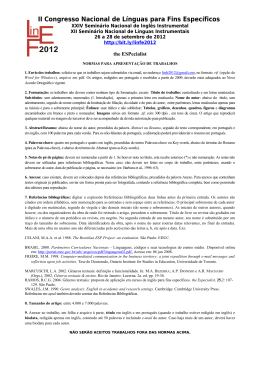
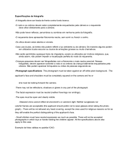
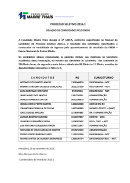
Eduardo Vedovatto
Movimentação dos dentes artificiais em próteses
totais com diferentes profundidades de palato.
Araçatuba
2009
Livros Grátis
http://www.livrosgratis.com.br
Milhares de livros grátis para download.
Eduardo Vedovatto
MOVIMENTAÇÃO DOS DENTES ARTIFICIAIS EM PRÓTESES
TOTAIS COM DIFERENTES PROFUNDIDADES DE PALATO.
Tese apresentada à Faculdade de Odontologia do Câmpus de
Araçatuba, Universidade Estadual Paulista “Júlio de Mesquita
Filho” – UNESP, como parte integrante dos requisitos para
obtenção do título de doutor, do programa de pós-graduação em
Odontologia, na área de Prótese Dentária.
Orientador: Prof. Titular Humberto Gennari Filho
Araçatuba
2009
Catalogação-na-Publicação
Serviço Técnico de Biblioteca e Documentação – FOA / UNESP
V416c
Vedovatto, Eduardo
Movimentação dos dentes artificiais em próteses totais
com diferentes profundidades de palato / Eduardo Vedovatto.
- Araçatuba : [s.n.], 2009
73 f. : il. ; tab. + 1 CD-ROM
Tese(Doutorado) – Universidade Estadual Paulista, Faculdade
de Odontologia, Araçatuba, 2009
Orientador: Prof. Humberto Gennari Filho
1. Prótese total 2. Resinas acrílicas 3. Dente artificial
Black D3
CDD 617.69
Dados Curriculares
Dados Curriculares
Eduardo Vedovatto
Eduardo Vedovatto nasceu em Valinhos – SP no dia 25 de maio de 1979. O primogênito
de Francisco de Assis Vedovatto e Aparecida Donizetti da Silva Vedovatto ingressou, após o
ensino fundamental, no Colégio Técnico de Campinas da Universidade Estadual de Campinas no
período de 1994 à 1997. Além de cumprir o ensino médio, também concluiu o curso de técnico
de mecânica se desbravando em seu primeiro estágio nas indústrias Kärcher Ltda (1997). Sua
carreira que parecia sedimentada em Engenharia Mecânica, sofreu uma guinada em 1998, ano
que decidira prestar para a área da saúde. Diferentemente de muitos que escolheram a
Odontologia como refúgio para a Medicina, Eduardo permaneceu firme em sua escolha mesmo
tendo sido aprovado na Faculdade de Medicina de Jundiaí em 1999. No mesmo ano, ingressou na
Faculdade de Odontologia de Araçatuba da Universidade Estadual Paulista. Adotou AraçatubaSP como segundo Lar, concluindo o curso de graduação em Odontologia em 2002. Dando
continuidade às atividades, realizou estágio no Departamento de Materiais Odontológicos e
Prótese em 2003 e em 2004 foi aprovado no mestrado do programa de pós-graduação em
Odontologia (área de Prótese Dentária) da mesma instituição. Como conseqüência, obteve em
2006 o título de Especialista em Prótese Dentária pelo Conselho Federal de Odontologia, mesmo
ano em que iniciou o Doutorado em Prótese Dentária pelo mesmo Programa. Nesse período
apresentou mais de 60 trabalhos científicos, foi autor e co-autor de 30 trabalhos (15 publicados),
e ganhou 4 prêmios. Complementarmente se aperfeiçoou em Periodontia (2002), em Estética e
Cosmética (2005) e realizou um MBA em gestão e markenting na saúde (2005). Atualmente
dirige a Clinica Vedovatto de Odontologia Contemporânea.
Dedicatória
Dedicatória
Este trabalho é totalmente dedicado em memória à
Maria por ter sonhado comigo doutor.
Agradecimentos especiais
Agradecimentos especiais
Aos meus pais pelo amor incondicional
À Cristina pelo amor que move meu coração
À Bianca pelo amor que teremos
À Erica pelo amor que nascemos
Ao Prof Humberto Gennari Filho pelo amor acadêmico.
Agradecimentos
Agradecimentos
À Faculdade de Odontologia de Araçatuba – UNESP que levarei para aonde for, pra
sempre!
Aos Professores e Amigos ou Amigos e Professores Marcelo Coelho Goiato e Eduardo
Piza Pellizzer. Obrigado pelo estímulo mesmo de baixo de chuva.
À Prof Dra Maria Cristina Rosifini Alves Resende por ter superado minhas expectativas
na ajuda de meu EGQ.
Aos amigos e grandes profissionais, José Vitor Quinelli Mazaro, Fellippo Ramos Verri e
Ricardo Shibayama por tudo o que fizeram por mim na profissão ou na vida.
À minha casa, o Departamento de Matérias Odontológicos e Prótese e todos que o
representam. Sentirei saudades!
Epígrafe
Epígrafe
“Na viagem de um lado ao outro na ordem da vida, nós aprendemos uma lição, que a natureza
viva não é um mecanismo, mas um poema”
Thomas H. Huxley, 1856
Sumário
SUMÁRIO
RESUMO
9
ABSTRACT
12
LISTA DE FIGURAS
15
LISTA DE TABELAS
16
LISTA DE QUADROS
18
1 Introdução
19
2 Materiais E Métodos
22
3 Resultados
30
4 Discussão
36
5 Conclusão
41
Referências Bibliográficas
43
ANEXOS
46
RESUMO
Resumo
Vedovatto, E. Movimentação dos dentes artificiais em próteses totais com diferentes
profundidades de palato [Tese]. Araçatuba: Universidade Estadual Paulista, 2009.
RESUMO
D
emonstração do problema: alterações oclusais sempre ocorrem durante o processamento
de próteses totais, entretanto, o deslocamento do dente no sentido ocluso-cervical e sua
relação com a profundidade do palato ainda não foi totalmente evidenciada.
Objetivos: esse estudo investigou a movimentação dos segundos molares superiores em próteses
totais processadas com resina acrílica, com diferentes profundidades de palato e com diferentes
materiais de inclusão.
Métodos: vinte e oito próteses totais superiores idênticas foram confeccionadas sobre o modelo
com palato raso e vinte e oito próteses sobre o modelo com palato profundo. Essas próteses foram
divididas em quatro grupos com 14 unidades de acordo com o tipo de palato e o tipo de material
de inclusão (gesso ou silicone). Foram demarcados pontos na face distal dos segundos molares e
no modelo como referência para a mensuração do deslocamento dos dentes. O deslocamento
vertical, horizontal e angular foi verificado através das imagens das próteses com o programa
AutoCad em dois momentos: antes da inclusão em mufla e depois da demuflagem (sem a
separação do modelo). Os valores foram submetidos ao tratamento estatístico de KolmogorovSmirnov e ANOVA com o Tukey e Fisher post hoc test a 5% de significância.
Resultados: Os resultados não mostraram diferença estatística significante entre os grupos nos
deslocamentos angular (p=0,674) e horizontal (p=0,856). Entretanto, no sentido cervico-oclusal,
foi observada diferença entre os grupos (p=0,012) apontando maiores alterações para aqueles cuja
inclusão foi realizada com o silicone (E3=0,047 e E2=0,044 cm)em relação ao gesso (E1 = 0,027
Resumo
e C= 0,026cm). Os grupos com diferentes profundidades de palato não apresentaram diferença
estatística significante entre si.
Conclusão: esse estudo pôde concluir que o tipo de palato não influenciou na movimentação dos
segundos molares, já o método de inclusão influenciou de maneira significativa no deslocamento
vertical desses dentes.
Implicação clínica: O conhecimento do modo como ocorre a movimentação dos dentes em
profundidades de palato diferentes e sua interação com os materiais de inclusão poderá melhorar
o desenvolvimento de técnicas que minimizem ou corrijam essas alterações e como conseqüência
próteses mais fiéis com maior desempenho e menor tempo de ajuste.
PALAVRAS-CHAVE: Prótese Total, Resinas Acrílicas, Dente Artificial.
ABSTRACT
Abstract
Vedovatto, E. Artificial tooth movement on complete dentures with different palate depth. [Tese].
Araçatuba: Universidade Estadual Paulista, 2009.
ABSTRACT
S
tatment of the problem: occlusal changes always occur during the complete dentures,
however, the cervical-occlusal displacement of the tooth and its relationship with the
plate depth has not yet been well clarified.
Purpose: This study investigated the movement of the second molars in complete dentures
processed with different palate depths and different materials for inclusion.
Methods: Twenty-eight complete maxillary dentures were made on cast with shallow palate and
twenty-eight on the cast with deep palate. These grafts were divided into four groups with 14
units according to the type of palate and the type of material for inclusion (gypsum or silicone).
Points were marked on the distal face of second molars and in the model for measuring the
displacement of the teeth. The vertical displacement, horizontal and angular was verified by the
images of the prosthesis with the AutoCad program on two occasions: before inclusion and after
deflasking (without the separation of the cast). The values were submitted to statistical
Kolmogorov-Smirnov test and ANOVA with Tukey's and Fisher's post hoc test at 5%
significance.
Results: The results showed no statistically significant difference between groups in the angular
displacement (p = 0674) and horizontal (p = 0856). However, the cervical-occlusal direction
(vertical displacement), difference was observed between the groups (p = 0012) indicating major
changes to those whose inclusion was performed with silicone (E3 = 0047 and E2 = 0044cm) on
gypsum (E1 = 0027 and C = 0026cm). The groups with different depths of the palate showed no
statistically significant difference between them.
Abstract
Conclusion: This study could conclude that the type of palate did not influence the movement of
the second molars, as the method of inclusion of a significant influence on the vertical
displacement of the teeth.
Clinical implications: The knowledge of how the movement of the teeth occurs at different
depths of the palate and its interaction with the material for inclusion may improve the
development of techniques that minimize or correct these changes and consequently more loyal
prostheses in less time for adjustment.
KEYWORDS: Complete Dentures, Acrylic Resins, Artificial Teeth.
Lista de Figuras
LISTA DE FIGURAS
FIGURA 1
Método de replicação das próteses. Dentes posicionados.
Note a área de escape para a cera (seta).
25
FIGURA 2
Prótese encerada sobre o palato raso (esquerda) e prótese
encerada sobre o palato profundo (direita).
26
FIGURA 3
Pontos demarcados na superfície distal e evidenciados com
grafite. Ponto transferido no modelo (região da tuberosidade)
e demarcado com grafite (seta).
26
FIGURA 4
Preenchimento total do ponto analisado em microscópio (50
vezes).
26
FIGURA 5
Imagem sendo mensurada no AutoCad 2005. Distância
horizontal (0,179) distância vertical (0,76) e distância
angular (98,145°).
29
FIGURA 6
Alterações nas três variáveis estudadas entre os grupos
35
Lista de Tabelas
LISTA DE TABELAS
TABELA 1
Análise de Variância fator duplo para o deslocamento
angular.
31
TABELA 2
Comparação pelo teste de Tukey para a interação.
31
TABELA 3
Teste de Tukey comparando somente os grupos.
32
TABELA 4
Teste de Tukey comparando o lado de análise para o
deslocamento angular.
32
TABELA 5
Análise de Variância fator duplo para o deslocamento
Horizontal.
32
TABELA 6
Teste de Tukey para a interação no deslocamento horizontal
33
TABELA 7
Teste de Tukey para o deslocamento comparando somente os 33
grupos.
TABELA 8
Teste de Tukey comparando o lado de análise para o
deslocamento Horizontal.
33
TABELA 9
ANOVA para o deslocamento vertical
34
TABELA 10
Teste de Fisher para a interação no deslocamento vertical.
34
TABELA 11
Teste de Fisher para a variável “grupo” considerando o
deslocamento vertical.
34
TABELA 12
Teste de Fisher para o local de análise considerando o
deslocamento vertical.
35
TABELA 13
Distribuição dos dados da análise vertical
49
TABELA 14
Teste de Kolmogorov-Smirnov para análise vertical
49
TABELA 15
Distribuição dos dados da análise horizonta
49
Lista de Tabelas
TABELA 16
Teste de Kolmogorov-Smirnov para análise horizontal
49
TABELA 17
Distribuição dos dados da análise angular
50
TABELA 18
Teste de Kolmogorov-Smirnov para análise angular
50
Lista de Quadros
LISTA DE QUADROS
QUADRO 1
Descrição resumida dos grupos estudados
24
INTRODUÇÃO
Introdução 20
1 INTRODUÇÃO
O
sucesso da terapia protética para o edentulismo total requer excelência técnica do
profissional e do técnico de laboratório. Com o passar dos anos, os pacientes tem se
beneficiado do melhoramento das técnicas de reabilitação, passando do método convencional,
para os métodos assistidos por implantes osseointegráveis. O manejo fisiológico e psicológico
dos pacientes desdentados tem auxiliado milhares de profissionais a reconsiderar as soluções
reabilitadoras por próteses totais removíveis. Como a análise da excelência estética, a boa
disposição dos dentes artificiais e a compensação gengival, tornam o tratamento com próteses
totais em recuperador facial. O domínio da oclusão através da determinação de equilíbrio e
balanceamento seguidos de bases bem adaptadas possibilitou a maximização do tratamento do
edentulismo, entretanto é muitas vezes insuficiente para atingir a necessidade do paciente. Um
importante critério para o sucesso da prótese total é a boa oclusão que será alcançada por meio da
individualização da mesma ainda em cera1,2 . Entretanto, o processamento das próteses reflete-se
na alteração do padrão oclusal, de modo que, em algumas vezes, a correção dessas alterações por
desgaste afeta seu desempenho clínico3.
A discrepância existente entre a oclusão com a prótese em cera e finalizada está
relacionada, principalmente com o processamento laboratorial4. Essas alterações podem não estar
somente relacionadas com a contração de polimerização da resina acrílica, havendo uma gama de
variáveis relacionadas4,5. Alguns estudos6, 7 atribuíram essas alterações à técnica de inclusão em
mufla, sendo as mais comuns realizadas pelo método convencional com gesso ou barreira de
silicone e gesso. Outros estudos4,8,9 responsabilizaram o método de processamento da resina
acrílica, se em banho de água quente, se microondas ou associados ao método de injeção.
Introdução 21
Embora esses fatores possam ser relevantes, foi assumido que a direção de movimentação
dos dentes artificiais pudesse estar relacionada com a geometria da base de resina acrílica10, 11, 12.
Hipoteticamente alguns autores10, 12, 13 relatavam uma diferença no padrão de contração da resina
sobre modelos com diferenças na profundidade e forma do palato, entretanto, não encontraram
diferença com significância estatística quando foi avaliada a desadaptação da base de resina.
Outra hipótese testada foi se a oclusão sofreria alterações influenciadas pela resina polimerizada
sobre diferentes formatos de palatos10 . Como resultados, analisados através de mensurações entre
os dentes no plano horizontal, o estudo indicou que alterações significantes, embora outro
estudo11 tivesse verificado que o tamanho da maxila não influenciou estatisticamente na
movimentação dos dentes artificiais no plano horizontal (entre os dentes).
O modo de como são avaliadas as alterações podem influenciar significativamente a
interpretação dos resultados4. A literatura traz inúmeros dados de alterações observadas no plano
oclusal e no reflexo sobre a alteração dimensional 1-8, 10, 11. Até o momento, o comportamento que
o dente pode sofrer no plano vertical não está bem esclarecido. Outro fator importante é o método
de mensuração. Microscópio comparador, compasso e perfilômetros são os meios mais utilizados
na literatura2,3,5,6,7. Outro método recente é o por computação gráfica4,
11
, que proporciona
maiores recursos para a comparação da movimentação dos dentes com confiabilidade.
Em decorrência da importância da oclusão para as próteses totais, esse estudo teve como
objetivo, verificar em três direções a alteração dos segundos molares em relação ao modelo,
quando as próteses totais superiores foram processadas com diferentes profundidades de palato e
diferentes métodos de inclusão. A hipótese nula (H0) aceita que as diferentes profundidades bem
como o método de inclusão não influenciam na quantidade de alteração dos dentes. A hipótese
alternativa (Ha) aceita que pelo menos uma das variáveis gera diferença na movimentação dos
dentes artificiais.
MATERIAL E
MÉTODOS
Material e Métodos 23
2 MATERIAL E MÉTODOS
E
sse estudo foi realizado comparando a movimentação dos segundo molares superiores em
relação ao modelo gesso por meio de computação gráfica. As mensurações foram
realizadas sobre as imagens das próteses capturadas antes do processamento (na fase de cera) e
após o processamento da resina acrílica (sem separar do modelo). Foi observado através da vista
posterior da prótese, a alteração de posição do molar em relação a inclinação do longo eixo, do
deslocamento vestíbulo/lingual e do deslocamento ocluso/cervical. As variáveis testadas foram as
diferentes profundidades de palato (raso ou profundo) bem como as diferentes técnicas de
inclusão (gesso ou barreira de silicone).
Os grupos testados
Quatro grupos foram avaliados (Quadro 1). O grupo que foi composto pelas próteses
confeccionadas sobre o modelo com características de palato raso e inclusão somente em gesso
foi considerado grupo controle (C). No primeiro grupo experimental (E1), as próteses foram
confeccionadas sobre o modelo com características de palato profundo e inclusão somente em
gesso. No segundo grupo experimental (E2), as próteses foram confeccionadas sobre o modelo
com palato raso e inclusão pela técnica da barreira de silicone. No terceiro e último grupo
experimental (E3), as próteses foram confeccionadas sobre o palato profundo e incluídas em
barreira de silicone. Baseado nos dados de estudo piloto, foi determinada um tamanho amostral
de 14 unidades para cada grupo, totalizando 56 amostras.
Material e Métodos 24
Quadro 1 – Descrição resumida dos grupos estudados
Grupo
Descrição
C
E1
E2
E3
modelo com palato raso e inclusão em gesso - rg
modelo com palato profundo e inclusão em gesso - pg
modelo com palato raso e inclusão em silicone - rs
modelo com palato profundo e inclusão em silicone - ps
Preparação das unidades experimentais
Um modelo de maxila desdentada foi selecionado para esse estudo. A face posterior do
modelo foi planificada em um recortador de gesso, para evidenciar o perfil do contorno posterior
do palato. Na região da tuberosidade foi realizada uma perfuração em formato cônico 1x1mm em
cada lado da tuberosidade, para que pudesse formar um segmento imaginário que serviu de
referência para as mensurações. Este modelo foi replicado com silicone para duplicação (Silibor
– Artigos Odontológicos Clássico Ltda, Brasil) para constituir 28 modelos idênticos, com
características de palato raso, inclusive com as duas demarcações posteriores11. Para o palato
profundo, um modelo com palato raso foi duplicado e a profundidade do palato foi alterada
através de cuidadoso desgaste para se obter um palato 10mm mais profundo e com formato em
“V”14. Cabe ressaltar que a crista do rebordo não foi alterada. Esse modelo modificado foi então
replicado pela técnica descrita para se obter os 28 modelos com palato profundo (n=28).
Uma prótese total foi encerada sobre o modelo com palato raso, respeitando uma
espessura de base de aproximadamente 2,5mm e a montagem dos dentes realizada em acordo
com a crista do rebordo12 . Como referência oclusal foi utilizada uma placa de vidro, que deveria
tocar a superfície oclusal e incisal dos dentes artificiais (montagem sem curva de compensação).
Material e Métodos 25
Para a replicação do posicionamento dos dentes bem como do enceramento, foi utilizada a
técnica da replicação (Figura 1) com silicone para duplicação4, 11.
O molde em silicone reproduziu o posicionamento dos dentes artificiais, o contorno da
base da prótese e a posição do modelo. Dentes artificiais idênticos (Vipi Dent plus 2D, 32M –
Dental Vipi Ltda, Pirassununga, Brasil) foram posicionados no molde seguidos pelo vazamento
de cera rosa 7 liquefeita (Wilson – Polidental Ind e Com Ltda, Brasil). Depois disso o modelo foi
posicionado no molde e após o endurecimento da cera, o modelo foi separado do molde obtendo
a cópia da prótese com a mesma posição dos dentes e as características de enceramento (figura
2).
Figura 1 – Método de replicação das próteses. Dentes
posicionados. Note a área de escape para a cera (seta).
Para o enceramento das próteses nos modelos com palato profundo, um index em silicone
foi realizado para transferir o mesmo posicionamento dos dentes obtidos na prótese com palato
raso. A forma de enceramento e espessura da base foram realizadas similarmente à prótese com
palato raso. Assim, vinte e oito próteses totais enceradas sob o palato profundo (Figura 2) foram
realizadas utilizando a técnica de duplicação em silicone já descrita.
Material e Métodos 26
Figura 2 – Prótese encerada sobre o palato raso (esquerda) e prótese
encerada sobre o palato profundo (direita).
Com as próteses em cera, uma delas foi eleita para se realizar a primeira demarcação nos
segundos molares. Dois pontos de aproximadamente 0,5 mm de diâmetro foram fisicamente
demarcados na face distal do segundo molar direito e esquerdo. Esses pontos formaram um
segmento de reta para tornar possível a mensurações (Figura 3). Um guia em resina acrílica, que
se apoiava sobre a oclusal dos dentes foi confeccionado para transferir a posição dos pontos para
as outras unidades experimentais. O método exigiu que todos os pontos fossem evidenciados com
grafite (Faber-Castell, Brasil) (Figuras 3 e 4). Portanto, para que não houvesse erro, toda extensão
foi preenchida e conferida em microscópio (Carl Zeiss – Citoval 2, Germany) com ampliação de
50 vezes (Figura 4).
Figura 3 – Pontos demarcados na superfície distal do
dente e evidenciados com grafite. Ponto transferido no
modelo (região da tuberosidade) e demarcado com
grafite (seta).
Figura 4 – Preenchimento total do ponto analisado
em microscópio (50 vezes).
Material e Métodos 27
Inclusão e processamento das próteses
O método de processamento eleito foi o processamento em microondas por ser prático e
cientificamente viável9,15, salientando que o mesmo pode interferir nos resultados, como
observado na literatura 8, 15, 16.
Portanto, muflas plásticas (modelo STG – Dental VIPI Ltda, Pirassununga, Brasil) foram
revestidas na base com gesso pedra tipo II (Herodent – Vigodent S/A, Brasil) para a fixação do
modelo, de modo que após a cristalização do gesso e isolamento do mesmo com vaselina (Ind.
Farmacêutica Rioquímica, São Paulo, Brasil), procedeu-se o revestimento da contramufla. Para
os grupos C e E1 a contramufla foi totalmente revestida com gesso pedra tipo III (HerodentVigodent S/A, Brasil) manipulados à vácuo (Turbomix – EDG equipamentos Ltda) conforme
instrução do fabricante. Já para os grupos E2 e E3, primeiramente as próteses foram revestidas
com uma barreira de silicone Labormass (Ruthinium, Ruthibras, Brasil) de aproximadamente 2
mm e posicionadas com pressão bidigital. Após a polimerização do silicone e da confecção de
retenções, gesso pedra tipo III (Herodent-Vigodent S/A, Brasil) foi utilizado para completar o
revestimento da contramufla.
Após a eliminação da cera, primeiramente em forno de microondas 2 min e lavadas com
água quente (100°C), as muflas foram secas em temperatura ambiente por 2 horas. A resina
acrílica de eleição foi a Vipi-wave (Lote 4123, rosa médio – Dental VIPI Ltda, Pirassununga,
Brasil) na proporção 2:1, manipulada até atingir a fase plástica (conforme recomendação do
fabricante) e prensadas uma única vez até atingir gradualmente 1250kgf, mantendo constante por
5min. Após a polimerização de bancada o processamento da resina ocorreu de acordo com o
fabricante em forno de microondas doméstico (Piccolo – Panasonic, Brasil) por 20 min a 160W e
5 min a 600W. Após o resfriamento em bancada por 24h16 as próteses foram demufladas e não
Material e Métodos 28
separadas do modelo. Os pontos foram limpos e imediatamente verificados no microscópio para
garantir o preenchimento com grafite.
Mensuração por computação gráfica
A mensuração dos ângulos bem como das distâncias dos molares foram realizadas em
imagens capturadas4, 11 e não diretamente nas próteses (Figura 5). Para a captura das imagens, a
superfície posterior do conjunto prótese/modelo foi posicionada paralelamente à superfície de um
scanner de mesa em conjunto com um bloco metálico com dimensões 10 x 10 x 10 mm. Para a
padronização, as imagens foram capturadas com 600ppi, área da imagem de 60 x 55 mm e o
bloco metálico sempre paralelo à mesa de luz do scanner (referência horizontal). As imagens
foram capturadas de cada unidade experimental após o enceramento dos dentes e após o
processamento da resina, representando estado inicial e final respectivamente4,11.
A inclinação bem como as distâncias dos segundos molares superiores em relação à base
do modelo foram mensuradas na imagem com o programa AutoCad 2005 (Autodesk Inc USA)
R16 versão N.63.0. As imagens foram exportadas uma a uma pelo comando ‘RASTER IMAGE`.
Esse comando possibilita trabalhar com imagens bitmaps em arquivos vetoriais. Portanto, a
imagem serviu como plano de fundo para as mensurações tendo o bloco metálico como um
padrão para determinar a escala da imagem11. Os pontos das imagens foram ampliados pelo
comando `ZOOM`, podendo traçar segmentos exatamente do centro de um ponto à outro. Após
realizar os traçados-guia, o comando `DIMENSION TOOLBAR` possibilitou medir as distâncias
dos pontos ou os ângulos entre os segmentos. As mensurações verticais, horizontais e angulares,
foram repetidas 3 vezes em cada imagem, sendo sua média considerada para este estudo. Depois
das mensurações, um local foi aleatoriamente selecionado para uma nova mensuração por dois
Material e Métodos 29
auditores devidamente calibrados. Esses dados foram comparados pelo teste de correlação de
Pearson para compor a confiabilidade e reprodutibilidade do método (r=0,98).
Figura 5 – Imagem sendo mensurada no AutoCad 2005. Distância horizontal (0,179), distância vertical (0,76) e
distância angular (98,145°)
Tabulação dos dados e análise estatística
Como foram mensuradas três vezes, a média aritmética de cada imagem foi considerada
para a análise estatística. A média das distâncias após a polimerização foi comparada com a
média das distâncias antes da polimerização. A diferença dessa mensuração resultou no
deslocamento relativo da posição do dente artificial. A média dos segmentos foi comparada
estatisticamente pelo ANOVA fator duplo e pelos testes de Tukey e Fisher a 5 % de significância.
RESULTADOS
Resultados 31
3 RESULTADOS
A
nálise angular
A tabela 1 mostra o tratamento estatístico ANOVA para a análise angular, tanto do lado
direito como do lado esquerdo. De acordo com o teste de distribuição Kolmogorov-Smirnov test
foi confirmado distribuição NORMAL (D=0,049). Os números negativos indicam inclinação
(rotação) para vestibular. O teste indicou que as variáveis isoladamente não produziram efeito
sobre o tratamento, entretanto, sua interação resultou num risco de 95,5% de chance de aceitar a
hipótese alternativa como verdadeira.
Tabela 1 – Análise de Variância fator duplo para o deslocamento angular.
Fonte da
variação
Local
Grupo
Local*Grupo
GL
1
3
3
S.Q.
Q.M.
0,383
7,088
17,881
0,383
2,363
5,960
F
0,178
1,098
2,769
Pr > F
0,674
0,354
0,045
A tabela 2 indica que maioria das alterações aconteceu no sentido vestibular, sendo que
apenas uma ultrapassou 1°(um grau). Diferença estatisticamente significante foi encontrada entre
apenas 2 grupos, considerando a interação (p=0,045).
Tabela 2 – comparação pelo teste de Tukey para a interação.
Categoria
Local-d*Grupo-E2
Local-e*Grupo-E1
Local-e*Grupo-E3
Local-d*Grupo-C
Local-d*Grupo-E3
Local-e*Grupo-C
Local-e*Grupo-E2
Local-d*Grupo-E1
LS
média
0,647
-0,262
-0,427
-0,476
-0,605
-0,633
-0,700
-1,121
Grupos
A
A
A
A
A
A
A
B
B
B
B
B
B
B
Resultados 32
Em relação ao grupo controle nenhum grupo sofreu alterações estatisticamente
significantes (p=0,354).
Tabela 3 - teste de Tukey comparando somente os grupos.
LS
média
-0,026
-0,516
-0,555
-0,691
Categoria
E2 (Rs)
E3 (Ps)
C (Rg)
E1 (Pg)
Grupos
A
A
A
A
A tabela 4 mostra os resultados do teste de Tukey para a variável “local”. A variável não
produziu efeito sobre a comparação como relatado pela tabela 1 (p=0,674).
Tabela 4 – teste de Tukey comparando o lado de análise para o deslocamento angular.
LS
média
-0,389
-0,506
Categoria
D
E
Grupos
A
A
Análise horizontal
Os altos valores de P expostos na tabela 5 indicam a maior probabilidade de rejeitar a
hipótese de que alguma variável ou sua interação possa surtir efeito no tratamento (Ha)
Tabela 5 - Análise de Variância fator duplo para o deslocamento Horizontal.
Fonte da
variação
Local
Grupo
Local*Grupo
GL
1
3
3
S.Q
0,000
0,000
0,000
Q.M.
0,000
0,000
0,000
F
0,014
0,257
0,591
Pr > F
0,906
0,856
0,622
As tabelas 6, 7 e 8 mostram o baixo valor das alterações no sentido horizontal, não
chegando a 0,006 cm (60 µm).
Resultados 33
Tabela 6 – Teste de Tukey para a interação no deslocamento horizontal
Categoria
Local-d*Grupo-C
Local-e*Grupo-E2
Local-d*Grupo-E1
Local-e*Grupo-E1
Local-d*Grupo-E3
Local-e*Grupo-C
Local-e*Grupo-E3
Local-d*Grupo-E2
LS
média
0,006
0,005
0,004
0,003
0,003
0,003
0,000
-0,001
Grupo
A
A
A
A
A
A
A
A
Tabela 7 - teste de Tukey para o deslocamento comparando somente os grupos.
Categoria
C (Rg)
E1 (Pg)
E2 (Rs)
E3 (Ps)
LS
média
0,004
0,004
0,002
0,001
Grupo
A
A
A
A
Tabela 8 - teste de Tukey comparando o lado de análise para o deslocamento Horizontal.
Categoria
D
E
LS
média
0,003
0,003
Grupo
A
A
Análise vertical
As tabelas com os dados estatísticos seguem também para a análise vertical conforme
apresentado. Entretanto, como o teste de Kolmogorov-Smirnov não indicou distribuição normal,
o teste post hoc eleito foi o Fisher.
Pela tabela 9 pode-se observar que apenas a variável “grupo” apresentou dados que
possibilitam rejeitar a Hipótese nula e considerar a Hipótese alternativa (p=0,012)
Resultados 34
Tabela 9 – ANOVA para o deslocamento vertical
Fonte da
variação
Local
Grupo
Local*Grupo
GL
1
3
3
S.Q.
0,001
0,010
0,002
Q.M.
0,001
0,003
0,001
F
1,070
3,808
0,731
Pr > F
0,303
0,012
0,536
A tabela 10 mostra que as alterações dos deslocamentos verticais variaram de acordo com
o local de análise e em relação ao grupo, Sendo a alteração mais crítica chegando a 0,050 cm
(500µm).
Tabela 10 – Teste de Fisher para a interação no deslocamento vertical.
Categoria
Local-d*Grupo-E3
Local-e*Grupo-E2
Local-e*Grupo-E3
Local-d*Grupo-E2
Local-d*Grupo-E1
Local-d*Grupo-C
Local-e*Grupo-C
Local-e*Grupo-E1
LS
média
0,050
0,046
0,044
0,042
0,037
0,027
0,025
0,017
Grupo
A
A
A
A
A
A
B
B
B
B
B
B
C
C
C
C
A tabela 11 mostra diferença estatisticamente significante entre os grupos que foram
tratados com silicone em relação aos grupos que foram revestidos somente com gesso (Figura 6).
Tabela 11 – Teste de Fisher para a variável “grupo” considerando o deslocamento vertical.
Categoria
E3 (Ps)
E2 (Rs)
E1 (Pg)
C (Rg)
LS
média
0,047
0,044
0,027
0,026
Grupo
A
A
B
B
Como nos testes anteriores, a tabela 12 não encontrou diferença estatística que seja
significante com relação ao local de análise para a movimentação vertical.
Resultados 35
Tabela 12 – Teste de Fisher para o local de análise considerando o deslocamento vertical.
Categoria
D
E
LS
média
0,039
0,033
Grupo
A
A
0,05
0
0,045
-0,1
0,04
(cm)
0,03
-0,3
0,025
-0,4
0,02
-0,5
0,015
-0,6
0,01
-0,7
0,005
0
-0,8
C (RG)
E1 (PG)
E2 (RS)
E3 (PS)
Figura 6 – Alterações nas três variáveis estudadas entre os grupos
(graus)
-0,2
0,035
vertical
horizontal
Angular
DISCUSSÃO
Discussão 37
4 DISCUSSÃO
A
s condições de processamento das próteses impostas pelo confinamento em mufla geram
concentrações de tensões desiguais repercutindo em um maior deslocamento de alguns
dentes em relação a outros1. Considerando, entre outras, essa afirmação, este estudo buscou
interpretar a movimentação dos dentes artificiais por meio da análise dos segundos molares
superiores de três modos: a inclinação do molar, seu deslocamento horizontal e seu deslocamento
vertical. Todas três variáveis representam um significado clínico importante na boa oclusão. A
inclinação tem relacionamento direto com a manutenção da curva de compensação e o
balanceamento da oclusão, o deslocamento horizontal com o bom posicionamento no dente em
relação ao rebordo remanescente e o deslocamento vertical, com a dimensão vertical de oclusão2,
6, 11, 15
. Entretanto é o conjunto de todas que afeta a boa oclusão pré-estabelecida em cera, sendo
que
essas
alterações,
invariavelmente
geram
contatos
prematuros
deflectivos
que,
necessariamente requerem ajuste oclusal direto, ou até mesmo remontagem em articulador11. Em
estudo com 121 pacientes, a remontagem no articulador proporcionou quase o dobro de
performance mastigatória em relação aos pacientes que receberam próteses não ajustadas
previamente no articulador3. Entretanto, caso o ajuste necessário afete muito a topografia oclusal
dos dentes artificiais, poderá influir em sua eficiência triturante2.
Quanto à análise angular, os dados estatísticos não apresentaram diferença entre os
tratamentos aplicados, aceitando a hipótese nula (h0). Embora seja fato, os dados enriquecem o
estudo do comportamento das alterações. Foi observada em todos os grupos uma tendência do
dente girar no sentido vestibular, ou seja, abrir o arco dental. Como comentado hipoteticamente10,
esperava-se que as próteses com palato raso movimentassem menos no sentido vestibular do que
as próteses com palato profundo. Entretanto isso aconteceu apenas para o Grupo RS (-0,026°) em
Discussão 38
detrimento aos demais que apresentaram em média alterações de -0,5° (tabela 2). Analisando a
interação (local x grupo) essas alterações parecem aleatórias . Com relação ao lado de análise, o
comportamento esperado seria a simetria de alterações, entretanto, como pudemos observar na
tabela 4, essas alterações foram em média diferentes, porém sem significância estatística. Se
compararmos os dados entre as unidades experimentais, fica claro a alteração maior ora de um
lado, ora de outro dentro de um mesmo grupo (tabela 2). Isso demonstra ausência de simetria e
essa diferença pode ser atribuída ao empenamento da base, principalmente, pela energia potencial
formada no resfriamento e liberada no momento da demuflagem13,16.
Portanto, os resultados obtidos para análise da inclinação do molar são de difícil
comparação com os trabalhos apresentados na literatura 2, 5, 10, 11 , pois a maioria compara apenas a
distância de pontos na superfície oclusal4. Aceitou-se, hipoteticamente, nas condições desse
estudo, que uma inclinação de 1° (um grau) representa em um distanciamento da ponta de
cúspide de 0,03 cm. Portanto, caso os dois molares movimentassem 1° (um grau) para vestibular,
as cúspides mésio-palatinas se distanciariam em 0,06 cm (600µm). A literatura apresenta
movimentações que variam de 0,1 mm (100µm)11, 0,2 mm (200 µm)8 até 0,51mm (510 µm)10
entre os molares, analisando horizontalmente apenas.
A movimentação do corpo do dente no sentido horizontal mostrou um comportamento
mais previsível e podem ser clinicamente insignificantes. As alterações que não apresentaram
diferenças estatísticas entre si, giraram em torno de 0,001 à 0,004cm (tabela 7). Apesar de
clinicamente e estatisticamente insignificantes, os grupos cuja inclusão foi gesso apresentaram
maior alteração numérica que aqueles cujas próteses foram revestidas com silicone (figura 6).
Cabe ressaltar que a movimentação do corpo do dente no sentido horizontal, não significa
exatamente a movimentação da cúspide do dente. Como visto anteriormente, a inclinação do
dente pode representar mais clinicamente do que o deslocamento horizontal.
Discussão 39
Deste modo, as alterações cujos valores apresentaram maior grandeza foram no sentido
vertical. Sua principal conseqüência clínica é o aumento da dimensão vertical de oclusão. Essa
alteração pode ser explicada pelo aumento da espessura da base de resina em decorrência da
prensagem, bem como da contração da base durante sua polimerização, acarretando em
movimentação horizontal dos dentes posteriores, contato oclusal prematuro e “pin openning”2, 5,
11
.Neste estudo foi constatado que o material de inclusão possui maior interferência na
movimentação vertical do que a profundidade do palato. Outras comparações de técnicas de
inclusão, se com gesso ou silicone, limitaram-se em analisar apenas as alterações de dimensão
vertical ou de distâncias horizontais ao longo dos dentes artificiais4, 7. Os resultados desse
trabalho em que a avaliação ocorreu no sentido vertical mostraram alterações estatísticas
significantes para os dois grupos com barreira de silicone, em relação aos incluídos com gesso.
Comparando a grandeza dos resultados com estudos científicos anteriores4, 5, 10, 11, vê-se
uma redução na distorção dos dentes artificiais. Atenção é requerida neste momento, pois o fato
de analisar a movimentação apenas dos dentes ou de um conjunto de dentes não é indicativo de
que o desempenho protético seja melhor. Outros fatores são reconhecidos como tão importantes
quanto, sendo um deles a adaptação da base de resina 12,13. Cabe ressaltar que, embora o gesso
tenha apresentado resultados melhores para algumas situações, nenhuma técnica pode ser containdicada, devendo ser ponderado seus respectivos custos-benefícios.
Entretanto, alteração do padrão oclusal pode significar perda do desempenho clínico das
próteses, uma vez que muitos profissionais não competem em ajustar adequadamente os
aparelhos quando a discrepância oclusal é grande. Pior ainda são os contatos deflectivos que
resultam
em
desvio
mandibular,
tendo
como
conseqüência
clínica
a
desordem
temporomandibular. Associa-se a isso o tempo requerido para os ajustes e o acréscimo dos
Discussão 40
honorários profissionais, interferindo no caráter sócio-econômico que a prótese total removível
representa na odontologia atual.
Na odontologia contemporânea não se pode esquecer que as próteses totais são em muitos
casos assistidas por implantes, uma vez que resolvida a estabilidade das mesmas, o equilíbrio
oclusal é primordial, para garantir sobrevida do tratamento.
CONCLUSÃO
42
5 CONCLUSÃO
C
onsiderando suas limitações, os resultados permitiram concluir que a profundidade do palato
não influenciou na magnitude e na direção da movimentação dos segundos molares, entretanto, o
a movimentação vertical foi diferente quando variou-se o tipo de revestimento utilizado para a
inclusão, apontando melhores resultados para os grupos cuja inclusão ocorreu com o gesso, em
relação ao silicone..
REFERÊNCIAS
BIBLIOGRÁFICAS
Referências Bibliográficas 44
REFERÊNCIAS BIBLIOGRÁFICAS
1. Mahler DB. Inarticulation of complete denture processed by compression molding
technique. J Prosthet Dent 1951; 1: 551-559.
2. Basso MFM, Nogueira SS, Arioli-Filho JN. Comparison of the occlusal vertical
dimension after processing complete dentures made with lingualized balanced occlusion
and conventional balanced occlusion. J Prosthet Dent 2006; 96: 200-4.
3. Sidhaye AB, Master SB. Efficacy of remount procedures using masticatory performance
tests. J Prosthet Dent 1979; 42: 129-133.
4. Shibayama R, Gennari-Filho H, Mazaro JVQ, Vedovatto E, Assunção W. Effect of
Flasking and Polymerization Techniques on Tooth Movement in Complete Denture
Processing. J Prosthodont 2009; 18: 259-264.
5. Becker CM, Smith DE, Nicholls JI. The comparison of denture-base processing
techniques. Part II. Dimensional changes due to processing. J Prosthet Dent 1977; 37:
451-459.
6. Mainieri ET, Boone ME, Potter RH. Tooth movement and dimensional change of denture
base materials using two investment methods. J Prosthet Dent 1980; 44: 368-373.
7. Sinclair GF, Clark RKF. Dimesional change in dentures processed in silicone and stone
moulds. Eur J Prosthodont Rest Dent 2002; 10: 43-45.
8. Keenan PLJ, Radford DR, Clark RKF. Dimensional Change in complete dentures
fabricated by injection molding and microwave processing. J Prosthet Dent 2003; 89: 3744.
9. Nishi M. Curing of denture base resins with microwave irradiation with particular
reference to heat curing resins. J Osaka Dent 1968; 2: 23-40.
Referências Bibliográficas 45
10. Abuzar MA, Jamani K, Abuzar M. Tooth movement during processing of complete
dentures and its relation to palatal form. J Prosthet Dent 1995; 73: 445-449.
11. Gennari-Filho H, Alves LMN, Santos PH, Goiato MC, Vedovatto E, Shibayama
R.Análises de las alteraciones de la posición de los dientes artificiales de protesís totales
maxilares em función del tamaño del arco. Acta Odont Ven 2007; 45:
12. Laughlin GA, Eick JD, Glaros AG, Young L, Moore D. A comparison of palatal
adaptation in acrylic resin denture bases using conventional and anchored polymerization
techniques. J Prosthodont 2001; 10: 204-211.
13. Sadamori S, Ganefiyanti T, Hamada T, Arima T. Influence of thickeness and location on
the residual monomer content of denture base cured by three processing methods. J
Prosthet Dent 1994; 72: 19-22.
14. Johnson DL, Holt RA, Duncanson-Jr MG. Contours of the edentulous palate. J Am Dent
Assoc 1986; 113: 35-40.
15. Nelson MW, Kotwal KR, Sevedge SR. Changes in vertical dimension of occlusion in
conventional and microwave processing of complete dentures. J Prosthet Dent 1991; 65:
306-308.
16. Komiyama O, Kawara M. Stress relaxation of heat-activated acrylic denture base resin in
the mold after processing. J Prosthet Dent 1998; 79: 175-181.
ANEXOS
Anexos 47
Bibliografia Recomendada
ü CAMPOS, M. S.; CAVALCANTI, B. N.; CUNHA, V. P. P. Occlusal changes in
complete dentures processed by pack-and-press and injection-pressing techniques. Eur. J.
Prosthodont., v. 13, n. 2, p.78-80, Jun. 2005.
ü CURY, A. A. D. B.; RODRIGUES JUNIOR, A. L.; PANZERI, H. Resinas acrílicas
dentais polimerizadas por energia de microondas, método convencional de banho de água
e quimicamente ativada: propriedades físicas. Rev. Odontol. Univ. São Paulo, v. 8, n. 4,
p. 243-249, Out./Dez. 1994.
ü HEARTWELL, C. M. Jr.; The effect of tissue resiliency on occlusion in complete denture
prosthodontics. J. Prosthet. Dent., v. 34, n. 6, p. 602-604, Dec. 1975.
ü HEGDE, V.; PATIL, N. Comparative evaluation of the effect of palatal vault
configuration on dimensional changes in complete denture during processing as well as
after water immersion. Indian J. Dent. Res., v. 15, n. 2, p. 62-65, Apr./Jun. 2004.
ü HUTTON, B.; FEINE, J.; MORAIS, J. Is there an association between edentulism and
nutritional state? J. Can. Dent. Assoc., v. 68, n. 3, p. 182-187, Mar. 2002.
ü JOHN, J.; GANGADHAR, S. A.; SHAH, I. Flexural strength of heat-polymerized
polymethyl methacrylate denture resin reinforced with glass, aramid, or nylon fibers. J.
Prosthet. Dent., v. 86, n. 4, p. 424-427, Oct. 2001.
ü KIPPAX, A.; WATSON, C. J.; BASKER, R. M.; PENTLAND, J. E. How well are
complete dentures copied? Br. Dent. J., v. 185, n. 3, p. 129-133, Aug. 1998.
ü LAI, C. P.; TSAI, M. H.; CHEN, M.; CHANG, H. S.; TAY, H. H. Morphology and
properties of denture acrylic resins cured by microwave energy and conventional water
bath. Dent. Mater., v. 20, n. 2, p. 133-141, Feb. 2004.
Anexos 48
ü NISHIGAWA, G.; MATSUNAGA, T.; MARUO, Y.; OKAMOTO, M.; NATSUAKI, N.
Finite element analysis of the effect of the bucco-lingual position of artificial posterior
teeth under occlusal force on the denture supporting bone of the edentulous patient. J.
Oral Rehabil., v. 30, n. 6, p. 646-652, Jun. 2003.
ü POLYZOIS, G. L.; KARKAZIS, H. C.; ZISSIS, A. J.; DEMETRIOU, P. P. Dimensional
stability of dentures processed in boilable acrylic resins. A comparative study. J.
Prosthet. Dent., v. 57, n. 5, p. 639-649, May 1987.
ü SHINKAI, R. S.; HATCH, J. P.; RUGH, J. D.; SAKAI, S.; MOBLEY, C. C.;
SAUNDERS, M. J. Dietary intake in edentulous subjects with good and poor quality
complete dentures. J. Prosthet. Dent., v. 87, n. 5, p. 490-498, May 2002.
ü SWORDS, R. L.; LATTA, G. H.; WICKS, R. A.; HUGET, E. F. Periodic evaluation of
the occlusal vertical dimension of maxillary denture from the wax trial denture through 48
hours after polymerization. J. Prosthodont., v. 9, n. 4, p. 189-194, Dec. 2000.
ü TAKAYAMA, Y. Studies on the thinning of the upper acrylic resin complete denture
with the reinforced palate. (Part 2) Influence of the different palate forms on the thin plate
dentures. Shika Rikogaku Zasshi, v. 21, n. 53, p. 48-63, Jan. 1980.
ü THOMAS, C. J.; WEBB, B. C. Microwaving of acrylic resin denture. Eur. J.
Prosthodont. Rest. Dent., v. 3, n. 4, p. 179-182, Jun. 1995.
Anexos 49
Tabela 13 – Distribuição dos dados da análise vertical
Statistic
Mean
Variance
Skewness
(Pearson)
Kurtosis
(Pearson)
Data
0,036
0,001
Parameters
0,036
0,001
3,115
0,000
20,272
0,000
Tabela 14 – Teste de Kolmogorov-Smirnov para análise vertical
D
p-value
alpha
0,127
0,049*
0,05
*p<0,05 aceita-se que a distribuição não é normal
Tabela 15 – Distribuição dos dados da análise horizontal
Statistic
Mean
Variance
Skewness
(Pearson)
Kurtosis
(Pearson)
Data
0,003
0,000
Parameters
0,003
0,000
1,047
0,000
4,365
0,000
Tabela 16 - Teste de Kolmogorov-Smirnov para análise horizontal
D
p-value
alpha
0,105
0,162
0,05
*p>0,05 aceita-se que a distribuição é normal
Anexos 50
Tabela 17 – Distribuição dos dados da análise angular
Statistic
Mean
Variance
Skewness
(Pearson)
Kurtosis (Pearson)
Data
-0,447
2,245
Parameters
-0,447
2,245
0,175
1,269
0,000
0,000
Tabela 18 - Teste de Kolmogorov-Smirnov para análise angular
D
p-value
alpha
0,049
0,950
0,05
*p>0,05 aceita-se que a distribuição é normal
Anexos 51
2008 Guidelines for Preparing Manuscripts for
The Journal of Prosthetic Dentistry
Updated by the Editor’s Office of The Journal of Prosthetic Dentistry . Originally prepared by
the late Carl O. Boucher, DDS, and associates at The Ohio State University, College of Dentistry
and School of Journalism, Columbus, Ohio.
Anexos 52
Submission Guidelines
We are pleased that you are interested in writing an article for The Journal of Prosthetic
Dentistry . In publishing, as in dentistry, precise procedures are essential. Your attention to
and compliance with the following policies will help ensure the timely processing of your
submission.
Length of Manuscripts
Manuscript length depends on manuscript type. In general, research and clinical science
articles should not exceed 10 to 12 double-spaced, typed pages (excluding references,
legends, and tables). Clinical Reports and Technique articles should not exceed 4 to 5
pages, and Tips articles should not exceed 1 to 2 pages. The length of systematic reviews
is variable.
Number of Authors
The number of authors is limited to 4; the inclusion of more than 4 must be justified in
the letter of submission. (Each author’s contribution must be listed.) Otherwise,
contributing authors in excess of 4 will be listed after the references.
Formatting
All submissions must be typed and double-spaced. Print on only 1 side of the paper. Paper
dimensions should be 8.5 x 11 inches with 1-inch margins on all sides.
Hard Copy and Electronic Files
Please submit an electronic file of the text and tables on a CD. Microsoft Word is the
preferred word processing program. Without an electronic copy of the text and tables, we
cannot submit the manuscript to our review process. If photographic prints accompany
the text, high quality electronic illustrations must be submitted upon initial submission
( see pages 13-15 for more information).
Copyright Transfer
In accordance with the Copyright Act of 1976, all manuscripts must be accompanied by
the following statement signed by EACH author individually. (Two authors, two
statements; four authors, four statements, etc.) If a manuscript number has been assigned,
it should be included at the end of the statement.
Anexos 53
Copyright Transfer/IRB Approval/HIPAA Compliance Statement
The Editorial Council for the Journal of Prosthetic Dentistry
(Print AUTHOR’S NAME HERE)_________________________________ has submitted
an
originally
authored
article
entitled
“(TITLE
GOES
HERE)__________________________________________________________________
________________________________________________________________________
________________________" to The Journal of Prosthetic Dentistry owned by the
Editorial Council (the ‘‘JPD’’) for publication in the ‘‘Journal of Prosthetic Dentistry,’’
which is published by Elsevier Inc. (‘‘Publisher’’). In exchange for publication of the
Article, Author represents and warrants to the JPD and the Publisher, together with their
officers and directors, that the article delivered for publication (the ‘‘Article’’) is original
and does not infringe the patent, trademark, copyright, trade secret rights or other
proprietary rights of third parties (‘‘IP Rights’’). Author also represents that, except as
indicated below, Author has no financial interest or arrangement with any entity which
interest or arrangement might be perceived to bear on the objectivity of the Article, unless
that financial interest or arrangement has been disclosed in writing to the JPD. Author
further represents that the Article was created in compliance with the provisions of the
Health Insurance Portability and Accountability Act (HIPAA) of 1996, and that the Article
contains no ‘‘protected health information’’ as that term is defined by HIPAA.
Author further represents that the study was performed in compliance with Author’s
institution’s appropriate policies related to the use of animal and/or human subjects and
human-derived material (the ‘‘IRB’’).
Author agrees to indemnify Publisher and the JPD and to hold Publisher and the JPD
harmless from any claims made, and costs, attorneys’ fees, damages or license fees
incurred by Publisher or the JPD as a result of any claim of infringement of IP Rights, or
any violation of HIPAA or IRB compliance based in whole or in part on the Article.
Author has a financial interest or arrangement with each of the following entities:
________________________________________________________________
________________________________________________________________
Name (print)_______________________________ AUTHOR’S NAME PRINTED
HERE
Date:______________________
Name (signature) ____________________________ AUTHOR’S SIGNATURE HERE
Manuscript number, if assigned:
Anexos 54
Checklist for Initial Submission
Letter of submission •
Copyright transfer statement for each author. Name printed, then signed below. •
Conflict of interest and financial disclaimer statement, if applicable •
Permission to reproduce previously published material, if applicable •
Informed consent for patient photographs, if applicable •
CD containing electronic files of manuscript contents. •
Electronic image files (see Guidelines, pages 13-15) •
The submission on the CD should consist of the double-spaced manuscript, which should
include, in this order:
Title page with proper information for all authors
- Abstract
- Article proper
- References
- Tables
- Legends for illustrations
Submission Address
Send all manuscripts and/or make inquiries to:
Dr. Carol A. Lefebvre, Editor
or Catherine A. Prysiazny, Editorial Manager
The Journal of Prosthetic Dentistry
Medical College of Georgia
1120 15 St., AD 2943 th
School of Dentistry
Augusta, GA 30912-1255
Phone: (706) 721-4558
Fax: (706) 721-4571
E-mail: [email protected]
Anexos 55
Types of Articles
Articles are classified as one of the following: research/clinical science article, clinical
report, technique article, systematic review, or tip from our readers. Required sections for
each type of article are listed in the order in which they should be presented.
RESEARCH REPORT, CLINICAL STUDY
The research report should be no longer than 10-12 double-spaced, typed pages and be
accompanied by no more than 12 high-quality illustrations.
Abstract (approximately 250 words): Create a structured abstract with the
following subsections: Statement of problem, Purpose, Material and Methods,
Results, and Conclusion. The abstract should contain enough detail to describe the
experimental design and variables. Sample size, controls, method of measurement,
standardization, examiner reliability, and statistical method used with associated level
of significance should be described in the Material and Methods section. Actual
values should be provided in the Results section.
Clinical Implications: In 2-4 sentences, describe the impact of the study results on
clinical practice.
Introduction: Explain the problem completely and accurately. Summarize relevant
literature, and identify any bias in previous studies. Clearly state the objective of the
study and the research hypothesis at the end of the Introduction. Please note that, for
a thorough review of the literature, most (if not all references) should first be cited in
the Introduction and/or Material and Methods section.
Material and Methods: In the initial paragraph, provide an overview of the
experiment. Provide complete manufacturing information for all products and
instruments used, either in parentheses or in a table. Describe what was measured,
how it was measured, and the units of measure. List criteria for quantitative
judgment. Describe the experimental design and variables, including defined criteria
to control variables, standardization of testing, allocation of specimens/subjects to
groups (specify method of randomization), total sample size, controls, calibration of
examiners, and reliability of instruments and examiners. Statistical tests and
associated significance levels should be described at the end of this section.
Results: Report the results accurately and briefly, in the same order as the testing
was described in the Material and Methods section. For extensive listings, present
data in tabular or graphic form to help the reader. Describe the most significant
findings and trends. Text, tables, and figures should not repeat each other. Results
noted as significant must be validated by actual data and p-values.
Discussion: Discuss the results of the study in relation to the hypothesis and to
relevant literature. If the results do not agree with other studies and/or with accepted
opinions, state how and why the results differ. Agreement with other studies should
also be stated. Identify the limitations of the present study, and suggest areas for
future research.
Anexos 56
Conclusions: Concisely list conclusions that may be drawn from the research; do
not simply restate the results. The conclusions must be pertinent to the objectives and
justified by the data. In most situations, the conclusions are true for only the
population of the experiment. All statements reported as conclusions should be
accompanied by statistical analyses.
References: Select and format references in accordance with the guidelines on page
10.
Tables: Create tables in accordance with the guidelines on page 11. •
Legends for illustrations: Concisely describe each illustration without directly
duplicating the main text.
CLINICAL REPORT
The clinical report describes the author’s methods for meeting a patient treatment
challenge. It should be no longer than 4 to 5 double-spaced, typed pages and be
accompanied by no more than 8 high-quality illustrations. In some situations, the Editor
may approve the publication of additional figures if they contribute significantly to the
manuscript.
Abstract: Provide a short, nonstructured, 1-paragraph abstract that briefly
summarizes the problem encountered and treatment administered.
Introduction: Summarize literature relevant to the problem encountered. Include
references to standard treatments and protocols. Please note that most, if not all,
references should first be cited in the Introduction and/or Clinical Report section.
Clinical Report: Describe the patient, the problem with which he/she presented,
and any relevant medical or dental background. Describe the various treatment
options and the reasons for selection of the chosen treatment. Fully describe the
treatment rendered, the length of the follow-up period, and any improvements noted
as a result of treatment. This section should be written in past tense and in paragraph
form.
Discussion: Comment on the advantages and disadvantages of the chosen treatment,
and describe any contraindications for it. If the text will only be repetitive of previous
sections, omit the Discussion.
Summary: Briefly summarize the patient treatment. •
References: Select and format references in accordance with the guidelines on page
10. •
Legends for illustrations: Concisely describe each illustration without directly
duplicating the main text.
DENTAL TECHNIQUE
The dental technique article presents, in a step-by-step format, a unique procedure helpful
to dental professionals. It should be no longer than 4 to 5 double-spaced, typed pages and
be accompanied by no more than 8 high-quality illustrations. In some situations, the Editor
may approve the publication of additional figures if they contribute significantly to the
manuscript.
Abstract: Provide a short, nonstructured, 1-paragraph abstract that briefly
summarizes he technique.
Anexos 57
Introduction: Summarize relevant literature. Include references to standard
methods and protocols. Please note that most, if not all, references should first be
cited in the Introduction and/or Technique section.
Technique: In a numbered, step-by-step format, describe each step of the technique.
Put the text in command rather than descriptive form (e.g., “Survey the diagnostic
cast” rather than “The diagnostic cast is surveyed.”) Include citations for the
accompanying illustrations.
Discussion: Comment on the advantages and disadvantages of the technique,
indicate the situations to which it may be applied, and describe any contraindications
for its use. Avoid excessive claims of effectiveness. If the text will only be repetitive
of previous sections, omit the Discussion.
Summary: Briefly summarize the technique presented and its chief advantages. •
References: Select and format references in accordance with the guidelines on page
10. •
Legends for illustrations: Concisely describe each illustration without directly •
duplicating the main text.
Systematic Review
The author is advised to develop a systematic review in the Cochrane style and format.
The Journal is transitioning away from literature reviews to systematic reviews. For more
information on systematic reviews, please see www.cochrane.org. An example of a
systematic review in the Journal is: Torabinejad M, Anderson P, Bader J, Brown LJ, Chen
LH, Goodacre CJ, Kattadiyil MT, Kutsenko D, Lozada J, Patel R, Petersen F, Puterman I,
White SN. Outcomes of root canal treatment and restoration, implant-supported single
crowns, fixed partial dentures, and extraction without replacement: a systematic review. J
Prosthet Dent 2007 Oct;98(4):285-311.
Briefly, the systematic review consists of the following:
An Abstract - using a structured format. (Statement of problem, Purpose, Material and
methods, Results, Conclusion)
The text of the review - consisting of an introduction (background and objective), methods
(selection criteria, search methods, data collection and data analysis), results (description
of studies, methodological quality, and results of analyses), discussion, authors’
conclusions, acknowledgements and conflicts of interest. References should be peerreviewed and follow JPD
Guideline format (page 12).
Tables and figures, if necessary - showing characteristics of the included studies,
specification of he interventions that were compared, the results of the included studies, a
log of the studies that were excluded, and additional tables and figures relevant to the
review.
Anexos 58
TIPS FROM OUR READERS
Tips are brief reports on helpful or timesaving procedures. They should be limited to
2 authors, be no longer than 250 words, and include no more than 2 high-quality
illustrations. Place the procedure in a numbered, step-by-step format; place the text in
command rather than descriptive or passive form (e.g., “Survey the diagnostic cast”
rather than “The diagnostic cast is surveyed”).
Formatting Instructions
FIRST PAGE ARRANGEMENT TITLE PAGE
Please see the example provided on the following page.
Title: The title should define the study’s scope, content, and clinical significance.
Capitalize only the first letter of the first word. Do not underline the title or bold it.
Abbreviations or trade names should not be used in the title.
Authors: Directly under the title, type the names and degrees of the authors. List
academic degrees only . No fellowship designations, please.
Institution(s): Directly under the authors’ names, type their individual institutional
affiliations and the cities, states, and countries (if not the United States) in which
these institutions are located. If necessary, provide the English translation of the
name of the institution. Do not underline. If the authors are not affiliated with an
institution, please list the city, state, and country (if not the United States) in which
the authors live.
Presentation/support information and titles: If the research was presented before
an organized group, type the name of the organization and the location and date of
the meeting. If the work was supported by a grant or any other kind of funding,
supply the name of the supporting organization and the grant number. List the
academic titles (e.g., Assistant Professor) and departmental affiliations of all
authors.
Contact information: List the mailing address, business telephone, fax number,and
e-mail address of the author who will receive correspondence.
Anexos 59
Collagen tube containers in alveolar ridge augmentation
Robert K. Gongloff, DMD, and Richard Lee, DDS
School of Dentistry, University of California-San Francisco; Veterans Administration
Medical
Center, San Francisco, Calif
Supported by grant No. 9099-02 from the Veterans Administration.
Presented at the International Association of Oral and Maxillofacial Surgeons annual
meeting,
Vancouver, British Columbia, Canada, May 1995.
Chief, Oral and Maxillofacial Surgery, Veterans Administration Medical Center; and
Associate a
Clinical Professor, Department of Prosthodontics, UCSF School of Dentistry.
Former Resident, Department of Prosthodontics, UCSF School of Dentistry. b
Corresponding author:
Dr Richard K. Gongloff
Dental Service 160
Veterans Administration Medical Center
123 Main St.
San Francisco, CA 94121
Fax: 123- 456-7777
E-mail: [email protected]
Anexos 60
Acknowledgements (The authors thank…….)
[ NOTE: Acknowledgments should appear at the end of the title page, rather than in
the text of he manuscript.]
Anexos 61
ABSTRACT
The abstract must be typed on a page separate from the main text. •
The abstract should include no abbreviations or manufacturing information. •
MAIN TEXT
Headings
Headings should contribute to the clarity of the article and appear in appropriate
places to indicate a shift from one section to another (eg, Discussion to
Conclusions).The use of subheadings may be appropriate in the Material and
Methods section but is generally discouraged in the Results and Discussion. All
headings should be typed flush with the left margin. Main headings (eg,
MATERIAL AND METHODS) should be in capital letters; subheadings (eg,
Specimen preparation) should be in lowercase letters.
Identification of product and manufacturing information
Refer to products in generic terms. Immediately following the term, provide the
following information in parentheses: product name and manufacturer’s name, city,
state, and country (if not the United States). For example: “The impressions were
poured in type IV stone (Denstone; Heraeus Kulzer, South Bend, Ind) and related to
each other with a fast-setting vinyl polysiloxane occlusal registration material
(Correct VPS Bite Registration; Jeneric/Pentron Inc, Wallingford, Conn).” If the
same manufacturer is cited multiple times, the city and state/country are required
only in the first citation. Use generic drug names; trade names may be listed in
parentheses at the point of first mention.
Personal communications
Do not list a personal communication in the References. Instead, provide the
following information in the text in parentheses: the name of the person with whom
you communicated, his/her highest academic degree, whether the communication
was oral or written, and the date of the communication.
Abbreviations
If abbreviations are used, provide the expanded form upon first mention and
abbreviate thereafter [for example, fixed partial denture (FPD)].
REFERENCES
Acceptable references and their placement
Most, if not all, references should first be cited in the Introduction and/or Material
and Methods section. Only those references that have been previously cited or that
relate directly to the outcomes of the present study may be cited in the Discussion.
Anexos 62
Only peer-reviewed, published material may be cited as a reference. Manuscripts in
preparation, manuscripts submitted for consideration, and unpublished theses are
not acceptable references. Abstracts are considered unpublished observations and
are not allowed as references unless follow-up studies were completed and
published in peer-reviewed journals. References to foreign language publications
should be kept to a minimum (no more than 3). They are permitted only when the
original article has been translated into English. The translated title should be
cited and the original language noted in brackets at the end of the citation. Textbook
references should be kept to a minimum, as textbooks often reflect the opinions of
their authors and/or editors. The most recent editions of textbooks should be used.
Evidence-based journal citations are preferred.
Reference formatting
References must be identified in the body of the article with superscript Arabic
numerals. The complete reference list, double-spaced and in numerical order, should
follow the Conclusions section but start on a separate page. Only references cited in
the text should appear in the reference list. Reference formatting should conform to
Vancouver style as set forth in “Uniform Requirements for Manuscripts Submitted
to Biomedical Journals” (Ann Intern Med 1997;126:36-47). List up to six authors. If
there are seven or more, after the sixth author’s name, add et al . •
Abbreviate journal names per the Cumulative Index Medicus. A complete list of
standard abbreviations is available through the PubMed website:
http://www.ncbi.nlm.nih.gov/entrez/query.fcgi Format for journal articles: Supply
the last names and initials of all authors; the title of the article; the journal name;
and the year, volume, and page numbers of publication. Do not use italics, bold, or
underlining for any part of the reference. Put a period after the initials of the last
author, after the article title, and at the end of the reference. Put a semi-colon after
the year of publication and a colon after the volume. Issue numbers are not
used in Vancouver style .
Jones ER, Smith IM, Doe JQ. Uses of acrylic resin. J Prosthet Dent 1985;53:120-9.
Format for books: The most current edition must be cited. Supply the names and
initials of all authors/editors, the title of the book, the city of publication, the
publisher, the year of publication, and the inclusive page numbers consulted. Do not
use italics, bold, or underlining for any part of the reference.
Zarb GA, Carlsson GE, Bolender CL. Boucher's prosthodontic treatment for
edentulous patients. 11th ed. St. Louis: Mosby; 1997. p. 112-23.
Note: References should not be submitted in Endnotes. Endnotes formatting cannot
be edited by the Editorial Office or reviewers, and must be suppressed or removed
from the manuscript prior to submission.
Anexos 63
TABLES
Tables should be self-explanatory and should supplement, not duplicate the text.
Provide all tables at the end of the manuscript, after the figure legends (if present) or
reference list. There should be only one table to a page. Omit internal horizontal and
vertical lines. Omit any shading or color. Do not list tables in parts (eg, Table Ia, Ib,
etc.). Each should have its own number. Number the tables in the order in which
they are mentioned in the text. Supply a concise legend that describes the content of
the table. Create descriptive column and row headings. Within columns, align data
such that decimal points may be traced in a straight line. Use decimal points, not
commas, to mark places past the integer (eg, 3.5 rather than 3,5). In a line beneath
the table, define any abbreviations used in a table. If a table (or any data within it)
was published previously, give full credit to the original source in a footnote to the
table. If necessary, obtain permission to reprint from the author/publisher.
The tables should be submitted in Microsoft Word, WordPerfect, or RTF format.
Microsoft Word is preferred. If a table has been prepared in Excel, it should be
imported into one of the abovementioned formats prior to submission.
ELECTRONIC IMAGE SUBMISSION
Submitting your illustrations in electronic format allows more accurate and higher
quality reproduction of your work. The following guidelines must be carefully
followed.
File Type
Electronic files should be submitted as TIFF files. Figures should not be
submitted as Microsoft Word, Corel Draw, Harvard Graphics, PowerPoint, or
other presentation software format.
Color images should be in CMYK (Cyan/Magenta/Yellow/Black) color format
(color space) as opposed to RGB (Red/Green/Blue) color format.
The figures should be submitted to the Journal on a CD-ROM.
File Dimensions and Size
The figure dimensions must be a minimum of 4 × 6 inches; the figures should be of
standard dimensions.
The figures must be oriented correctly when submitted.
Figures should be size-matched (the same physical size), unless the image type
prohibits the
size-matching of the figure to other figures within the manuscript, as in the case of
panoramic or
Anexos 64
periapical radiographs, SEM images, graphs. Do not “label” the faces of the
figures with lette rs or numbers to indicate the order in which the figures should
appear, as such labels will be inserted during the publication process.
Clinical figures should be color balanced .The figures should be of professional
quality and high resolution. The following guidelines with respect to resolution
must be followed:
Color and black-and-white photographs should be created and saved at a
minimum of 300 dots per inch (dpi). (Note: A 4 × 6-inch image at a
resolution of 300 dpi will be approximately 6 megabytes, in terms of file
size.) A figure of less than 300 dpi must not be increased to 300 dpi; the
resulting quality and resolution will be poor.
• Line art should be created and saved as 1200 dpi.
• Combination artwork (an illustration containing both line art and photogra
ph) should be created and saved as 600 to 1000 dpi.
• All images must be easily readable and have good contrast. Clarity and
quality should be uniform among the parts of a multipart figure, and among
all of the figures within a manuscript. Composite figures (multiple images
combined into a single frame) are discouraged. Each image part should be a
separate 4 × 6-inch, 300-dpi image.
• A uniform background, preferably of a nontextured, medium blue, should
be provided for color figures when possible.
Line arest created in native design format, such as EPS t and combination artwork is
b(Encapsulated PostScript), Adobe Illustrator, InDesign, etc., but should be saved
as TIFF (Tagged Image File Format) files prior to submission to the Journal .
Color and bla ck-and-white photographs are best created and saved as TIFF images.
If a key to an illustration requires artwork (screen lines, dots, unusual symbols), it
should be incorporated into the drawing instead of included in the typed legend. All
symbols should be done professionally, be visible against the background, and be of
legible proportion should the illustration be reduced for publication. If text is to
appear within the figure, labeled and unlabeled versions of the figures must be
provided. Text appearing within the labeled versions of the figures should be in Ariel
font. The text should be sized to be easily read if the figure is reduced in size when
reproduced in the Journal. It is recommended that text no smaller than 10 point be
used. Lettering should be in proportion to the drawing, graph, or photograph. A
consistent font size should be used throughout each figure, and for all figures, to
ensure readability and a professional appearance.
Please note: Titles and captions should not appear within the figure file, but should be
provided in the manuscript text (see Figure Legends, below).
All microscopic photographs must have a measurement bar and unit of measurement
on the image. Color illustrations may be submitted when their use considerably
enhances the value of the manuscript. The Editor has final authority to determine
whether color illustrations provide the most effective presentation. Generally, a
maximum of figures will be accepted for clinical report and dental technique articles,
and 2 figures will be accepted for tips from our reader
Anexos 65
articles. However, the Editor may approve the publication of additional figures if they
contribute significantly to the manuscript.
In the article, clearly reference each illustration by including its number in
parentheses at the end of the appropriate sentence, before closing punctuation. For
example: “The sutures were removed after 3 weeks (Fig. 4).”
File Naming
Each figure mu st be numbered according to its position in the text (Figure 1, Figure
2, and soon), using Arabic numerals. The electronic image files must be named so
that the figure number and format can be easily identified. For example, a Figure 1 in
TIFF format should be named fig1.tif. Multipart figures must be clearly identifiable
by the file names: fig1A.tif, fig1B.tif, fig1C.tif, etc.
Figure Legend s
The figure legends should appear within the text of the manuscript, on a separate
page following the references and tables, and should appear under the heading
“LEGENDS.”
If an illustration is taken from previously published material, the legend must gi ve
full credit to the source (see Permissions).
Authors are obligated to disc lose whether illustrations have been modified in
any way.
Graphs
They should be numbered as figures and the fill for bar graphs should be distinctive
and solid; shading and patterns should be avoided. Thick, solid lines should be used,
and bold, solid lettering. Times New Roman font is preferred. Place lettering on
white background and avoid reverse type (white lettering on a dark background).
1200 dpi images should be provided if black and white.
The Journal reserves the right to standardize the format of graphs and tables.
PERMISSIONS
All quoted material must be clearly marked as such with quotation marks and a
reference number. If more than 5 lines are quoted, a letter of permission must be
obtained from the author and publisher of the quoted material.
If quotations are more than 1 paragraph in length, use open quotation marks at the
beginning of each paragraph and a closed quo tation mark the end of the final
paragraph only.
Anexos 66
Type all quoted material exactly as it appears in the original source, with no changes
in spelling or punctuation. Indicate material omitted from a quotation with ellipses
(3 dots for material omitted from within a sentence, 4 dots for material omitted after
the end of a sentence.)
If any submitted photos include the eyes of a patient, the patient must sign a consent
form authorizing use of his/her photo in the Journal. If such permission is not
obtained, the eyes will be blocked with black bars at publication. Illustrations that
are reprinted or borrowed from other published articles/books cannot be used
without the permission of the original author and publisher. The manuscript author
must secure this permission and submit it for review . In the illustration legend,
provide the full citation for the original source in parentheses
INTEREST IN COMMERCIAL COMPANIES AND OR PRODUCTS
Authors may not directly or indirectly advertise equipment, instruments, or
products in which they have a personal investment.
Statements and opinions expressed in the manuscripts are those of the authors and
not necessarily those of the editors or publisher. The editors and publisher disclaim
any responsibility or liability for such material. Neither the editors nor the publisher
guarantee, warrant, or endorse any product or service advertised in the Journal;
neither the editors nor the publisher guarantee any claim made by the manufacturer
of said product or service.
Authors must disclose any financial interest they may have in products mentioned in
an article. This disclosure should be typed after the Conclusions section.
Anexos 67
Writing Guidelines
GENERAL POLICIES AND SUGGESTIONS
Authors whose native language is not English should obtain the assistance of an
expert in English and scientific writing before submitting their manuscripts.
Manuscripts that do not meet basic language standards will be returned pre-review.
Do not use first person (I, we, us, our, etc.), which violates the objective tone
desired in scientific writing. “We conducted the study” can be changed easily to
“The study was conducted.” Describe experimental procedures, treatments, and
results in past tense. Describe teeth by name (eg, maxillary right first molar), not
number. It is generally better to paraphrase information from a published source
than to use direct quotations. Paraphrasing saves space. The exception is a direct
quotation that is unusually pointed and concise. When long terms with standard
abbreviations (as in TMJ for temporomandibular joint ) are used frequently, spell
out the full term upon first use and provide the abbreviation in parentheses. Use
only the abbreviation thereafter. Abbreviate units of measurement without a period
in the text and tables (eg, 9 mm). Use capital letters for proper names (persons,
institutions, etc.) and for proprietary product names but not for such terms as
profession , dentist , dentistry , and prosthodontics . Proprietary names function as
adjectives. Nouns must be supplied after their use, as in Vaseline petroleum jelly .
Wherever possible, use only the generic term.
OBJECTIONABLE TERMS
The following are selected objectionable terms and their proper substitutes. For a
complete list of approved prosthodontic terminology, consult the eighth edition of
the Glossary of Prosthodontic Terms (J Prosthet Dent 2005;94:10-92).
Incorrect
Alginate
Bite
Bridge
Case
Cure
Final
Freeway space
Full denture
Lower (teeth, arch)
Model
Modeling compound
Muscle trimming
Overbite, overjet
Correct
Irreversible hydrocolloid
Occlusion
Fixed partial denture
Patient, situation, or treatment as appropriate
Polymerize
Definitive
Interocclusal distance
Complete denture
Mandibular
Cast
Modeling plastic impression compound
Border molding
Vertical overlap, horizontal overlap
Anexos 68
Periphery
Post dam, postpalatal seal
Prematurity
Saddle
Study model
Upper (teeth, arch)
X-ray, roentgenogram
Border
Posterior palatal seal
Interceptive occlusal contact
Denture base
Diagnostic cast
Maxillary
Radiograph
In addition, sample is often used when specimen is meant.
CHECKLIST FOR EFFECTIVE WRITING
The article is long enough to convey the author's message—no shorter and no
longer. The author analyzes the audience and aims the message directly at that
audience. The material is well organized. The major divisions of the article are
distinct and follow each other in logical succession. The material within each
division flows smoothly. (Outlining is the key to success in this difficult task.)
The information is accurate. Even a minor discrepancy throws doubt on valid
portions of the article. The article is complete. It answers all the questions it raises;
it provides all the information it promises. The article is objective. Even opinion,
which is quite proper in the discussion, should be firmly rooted in fact. The article is
in a form that is as nearly final as the author and editor can make it before it is sent
to the publisher for formatting. It is expensive to make changes at the production
stage, so changes on the proof must be minor and absolutely necessary. The writing
is clear, concise, and grammatically correct. •
SOME ELEMENTS OF EFFECTIVE STYLE
Short words . Short words are preferable to long ones if the shorter word is equally
precise.
Familiar words . Readers want information that they can grasp easily and quickly.
Simple, familiar words provide clarity and impact.
Specific rather than general words . Specific terms pinpoint meaning and create
word pictures; general terms may be fuzzy and open to varied interpretations.
Brisk opening . Plunge into your subject in the first paragraph of the article. •
Limited use of modifying words and phrases . Check your adjectives, adverbs , and•
prepositional phrases. If they are not needed, strike them out.
No unnecessary repetition . An idea may be repeated for emphasis—so long as that
repetition is effective.
Short sentence length . No more than 20 words is recommended. Rambling
sentences, cluttered with subordinate clauses and other modifiers, are hard to read
and may cause.
Anexos 69
readers to lose their train of thought. Short sentences should, however, be balanced
with somewhat longer ones to avoid monotony.
Restraint . The writer who uses flamboyant words or overstates his proposition or
conclusions discredits himself. Facts speak f or themselves.
Clearly stated conclusions . Don’t hedge. If you don’t know something, say so.
Anexos 70
Additional Terminology Guidelines
Acrylic An adjective form that requires a noun, as in acrylic resin .
Affect, effect
Affect is a verb; effect is a noun.
African American Spelled thus and preferred over Negro and black in both
adjective ( African American patients ) and noun (of whom 20% were African
Americans ) forms.
Ampersand Should be avoided except in the name of a firm, as in John Smith &
Co.
Average, mean, Mean and average are synonyms. Median refers to the midpoint
in a range
median of items; the midpoint has many items above as below it.
Basic
Like fundamental , this word is often unnecessary. An example of
unnecessary use: Dental implants consist of two basic types: subperiosteal and
endosteal.
Between, among Use between when 2 things are involved and among when
there are more than 2.
Biopsy
This noun should NOT be used as a verb.
performed on the tissue rather than The tissue was biopsied.
Centric
An adjective that requires a noun, as in
A biopsy was
centric relation .
Currently, now, These expressions are often unnecessary, as in This technique is
at present, etc. currently being used . . .
Data
Use as a plural, as in
The data were . . .
Employ Should not become an elegant variation of use , as in This method is
employed . . .
Ensure Preferred over insure in the sense of to make certain .
Fad words This group includes the "ize" family (
conceptualize , prioritize ,
surgerize , finalize , etc.) and such terms as interpersonal , interrelationships ,
input , and viable . Several of these "words” have no dictionary standing at all
(even those that do should be used with caution).
Fewer, less Use fewer with nouns that can be counted ( fewer patients were seen
) and less with nouns that cannot be counted ( less material was used ).
Anexos 71
Following After is preferred.
Imply, infer
The speaker implies; the listener infers.
Incidence
The rate at which a disease occurs in a given time period; sometimes
confused with prevalence (the total number of cases of a disease in a given region).
Its, it's
Its is the possessive; it's is the contraction for it is .
Majority Means more than half ; use most when you mean almost all .
Male, female
and girls .
For adult humans, use
men and women . For children, use boys
Must, should
Must means that the course of action is essential. Should is less
strong and means that the course of action is recommended.
Numbers
Spell out numbers used in titles or headings and numbers at the
beginning of a sentence. The spelled version may also be preferable in a series of
consecutive numbers that may confuse the reader (eg, 2 3.5-inch disks should be
written two 3.5-inch disks ). In all other cases, use Arabic numerals.
Proper form; avoid orientate .
Orient
Paper (as in
manuscript)
Use article .
Pathologic
Use instead of
pathological . Other words in which the suffix –a
has been dropped include biologic, histologic , and physiologic .
Pathology
of disease)
The study of disease; often mistaken for
pathosis (the condition
Percent Use the percent sign in the text, as in The distribution of scores was as
follows: adequate, 8%; oversized, 23%; and undersized, 69%. But spell out when
the percent opens a sentence, as in Twenty percent of the castings
Principal, principle Principal is an adjective; it means main or chief , as in the
principal reason . Principle is a noun; it refers to a fundamental truth, standard, or
rule, as in the principles of Anglo-Saxon law.
Prior to
Before is preferred.
Rare, infrequent,
with a specific
Whenever possible, these vague terms should be backed up
Anexos 72
often not, etc. number.
Rather Like very , this word should be avoided.
Regimen A planned program for taking medication, dieting, exercising, etc. Not
to be confused with regime , meaning a system of government or management.
Symptomatology The science or study of symptoms; this word is not a synonym
for the word symptoms .
Technique Preferred over technic .
Utilize
Vertical
Very
Use is preferred.
An adjective that needs a noun, as in
vertical relation .
Frequently unnecessary; avoid.
Via Use through , with , or by means of .
White
Preferred over Caucasian . This is true only if the patient is from the
Caucasus region of Eastern Europe. If not, use the term, white to describe the
patient.
Anexos 73
Approved Abbreviations for Commonly Cited Journals
Because The Journal of Prosthetic Dentistry is published not only in print but also
online, authors must use the standard PubMed abbreviations for journal titles. If
alternate or no abbreviations are used, the references will not be linked in the online
publication. A complete list of standard abbreviations is available through the
PubMed website: http://www.ncbi.nlm.nih.gov/entrez/query.fcgi
Acta Odontologica Scandinavica
Acta Odontol Scand
American Journal of Orthodontics
Am J Orthod
Angle Orthodontist
Angle Orthod
British Dental Journal
Br Dent J
Cleft Palate Journal
Cleft Palate J
Dental Clinics of North America
Dent Clin North Am
Dental Digest
Dent Dig
Dental Practitioner and Dental Record
Dent Pract Dent Rec
Dental Progress
Dent Prog
Dental Survey
Dent Surv
International Dental Journal
Int Dent J
Implants International Journal of Oral and Maxillofacial Implants
Int J Oral
Maxillofac
International Journal of Periodontics and Restorative Dentistry Int J Periodontics
Restorative Dent
International Journal of Prosthodontics Int J Prosthodont
Journal of the American College of Dentists
J Am Coll Dent
Journal of the American Dental Association
J Am Dent Assoc
Journal of Dentistry for Children
J Dent Child
Journal of Dental Education
J Dent Educ
Journal of Dental Research J Dent Res
Journal of Endodontics
J Endod
Journal of Oral Rehabilitation
J Oral Rehabil
Journal of Oral Surgery
J Oral Surg
Journal of Periodontology
J Periodontol
Journal of Prosthetic Dentistry J Prosthet Dent
Journal of Prosthodontics
J Prosthodont
Oral Surgery, Oral Medicine, and Oral Pathology
Oral Surg Oral Med Oral
Pathol
Quintessence International
Quintessence Int
Livros Grátis
( http://www.livrosgratis.com.br )
Milhares de Livros para Download:
Baixar livros de Administração
Baixar livros de Agronomia
Baixar livros de Arquitetura
Baixar livros de Artes
Baixar livros de Astronomia
Baixar livros de Biologia Geral
Baixar livros de Ciência da Computação
Baixar livros de Ciência da Informação
Baixar livros de Ciência Política
Baixar livros de Ciências da Saúde
Baixar livros de Comunicação
Baixar livros do Conselho Nacional de Educação - CNE
Baixar livros de Defesa civil
Baixar livros de Direito
Baixar livros de Direitos humanos
Baixar livros de Economia
Baixar livros de Economia Doméstica
Baixar livros de Educação
Baixar livros de Educação - Trânsito
Baixar livros de Educação Física
Baixar livros de Engenharia Aeroespacial
Baixar livros de Farmácia
Baixar livros de Filosofia
Baixar livros de Física
Baixar livros de Geociências
Baixar livros de Geografia
Baixar livros de História
Baixar livros de Línguas
Baixar livros de Literatura
Baixar livros de Literatura de Cordel
Baixar livros de Literatura Infantil
Baixar livros de Matemática
Baixar livros de Medicina
Baixar livros de Medicina Veterinária
Baixar livros de Meio Ambiente
Baixar livros de Meteorologia
Baixar Monografias e TCC
Baixar livros Multidisciplinar
Baixar livros de Música
Baixar livros de Psicologia
Baixar livros de Química
Baixar livros de Saúde Coletiva
Baixar livros de Serviço Social
Baixar livros de Sociologia
Baixar livros de Teologia
Baixar livros de Trabalho
Baixar livros de Turismo
Baixar