SEBASTIÃO RODRIGO FERREIRA
ATIVIDADE OVICIDA DO FUNGO Pochonia chlamydosporia SOBRE OVOS DE
Ascaris suum E ATIVIDADE PREDATÓRIA DE FUNGOS NEMATÓFAGOS
SOBRE FORMAS INFECTANTES DE Oesophagostomum spp.
Dissertação apresentada à Universidade
Federal de Viçosa, como parte das
exigências do Programa de PósGraduação em Medicina Veterinária,
para obtenção do título de Magister
Scientiae.
VIÇOSA
MINAS GERAIS – BRASIL
2011
i ii
iii
__________________
O futuro pertence àqueles que acreditam na beleza de seus sonhos.
Elleanor Roosevelt
ii
AGRADECIMENTOS
A Deus, pelas bênçãos, cuidados e por fazer-me acreditar que nada é impossível.
A Universidade Federal de Viçosa, em particular ao Departamento de Veterinária pela
estrutura disponibilizada e o conhecimento que me foi ofertado.
Ao Conselho Nacional Desenvolvimento e Pesquisa (CNPq) pelo apoio financeiro, sem o
qual minha estadia em Viçosa teria sido mais árdua.
Ao Professor Jackson Victor de Araújo, pela oportunidade, orientação e amizade.
Aos meus pais, Braz e Maria Tereza, pelo apoio, amor e carinho.
Aos meus irmãos, cunhados, cunhadas e sobrinhos pelo apoio e torcida pela realização de
meus sonhos.
A minha namorada Michely, a quem conheci durante o mestrado, obrigado por entender
meus momentos de desatenção e hiperatividade, pelo carinho e companheirismo.
Aos amigos do laboratório: Juliana, Rogério, Luísa, Jose Geraldo (Tuim), Ademir, Tavela,
Fernanda, Manoel, Ingrid, Anderson, Layane, Luana e Wendeo, obrigado aos que, em
algum momento, contribuíram na realização deste trabalho e outros que durante nosso
convívio tornaram-no um momento de descontração e alegria.
A você amigo Fabio, muito obrigado pela amizade e pelos conselhos que foram cruciais na
realização deste trabalho.
Aos funcionários DVT, Rose, Beth, Izabel e Geraldinho, obrigado pela boa vontade e
presteza.
Ao Departamento de Zootecnia, de modo especial ao prof. Aluísio pela co-orientação e por
ter concedido o espaço e os animais para realização do experimento.
Ao prof. Leandro Grassi pela co-orientação e por ter cedido alguns isolados fúngicos de
Pochonia chlamydosporia
Aos funcionários da suinocultura do DZO, obrigado pela ajuda e receptividade.
iii
Aos companheiros de república, Henrique, Farley, Fred, Léo, Hilton, Lucas, obrigado pelo
convívio e, muitas vezes, pelos esclarecimentos e discussões que contribuíram para
construção do conhecimento.
A Fatinha, Wagner e demais amigos de Viçosa, obrigado pela amizade e pelos momentos
de descontração, vocês fizeram com que essa estadia em Viçosa fosse muito mais
prazerosa.
iv
BIOGRAFIA
Sebastião Rodrigo Ferreira, nasceu em 20 de janeiro de 1984, em Eugenópolis-MG. Filho
de Braz Lurde Ferreira e Maria Tereza Ferreira. Concluiu o ensino médio em 2001, na
Escola Estadual “Assis Brasil” – Vieiras, MG. No ano de 2004, iniciou o curso de
graduação em Ciências Biológicas, na Faculdade de Filosofia Ciências e Letras Santa
Marcelina, e graduou-se no ano de 2007. Em agosto de 2009, ingressou no mestrado no
programa de Pós-graduação em Medicina Veterinária da Universidade Federal de Viçosa
v
SUMÁRIO
1. RESUMO................................................................................................................xiii
2. ABSTRACT..............................................................................................................ix
3. Introdução Geral.........................................................................................................1
4. Capítulo 1...................................................................................................................5
Ovicidal activity of seven Pochonia chlamydosporia fungal isolates on Ascaris suum
eggs.
4.1 Abstract................................................................................................................6
4.2 Introduction.........................................................................................................6
4.3 Material and methods..........................................................................................7
4.4 Results.................................................................................................................8
4.5 Discussion..........................................................................................................10
4.6 References..........................................................................................................13
5. Capítulo 2.................................................................................................................16
Biological control of Ascaris suum eggs by Pochonia chlamydosporia fungus
5.1 Abstract..............................................................................................................17
5.2 Introduction........................................................................................................17
5.3 Material and methods.........................................................................................18
5.4 Results................................................................................................................20
5.5 Discussion..........................................................................................................22
5.6 References..........................................................................................................23
6. Capítulo 3.................................................................................................................27
Nematophagous fungi against infective larvae Oesophagostomum spp of pigs
6.1 Abstract..............................................................................................................28
6.2 Introduction .......................................................................................................28
6.3 Material and methods.........................................................................................29
6.4 Results and Discussion.......................................................................................31
6.5 References .........................................................................................................36
7. Capítulo 4.................................................................................................................38
vi
In vitro predatory activity of nematophagous fungi Duddingtonia flagrans on
infective larvae of Oesophagostomum spp. after passing through gastrointestinal
tract of pigs
7.1 Abstract...............................................................................................................39
7.2 Introduction........................................................................................................39
7.3 Material and methods.........................................................................................40
7.4 Results................................................................................................................41
7.5 Discussion...........................................................................................................42
7.6 References..........................................................................................................46
8. Conclusões................................................................................................................50
9. Referências bibliográficas .......................................................................................51
vii
RESUMO
FERREIRA, Sebastião Rodrigo, M.Sc., Universidade Federal de Viçosa, agosto de 2011.
Atividade ovicida do fungo Pochonia chlamydosporia sobre ovos de Ascaris suum e
atividade predatória de fungos nematófagos sobre formas infectantes de
Oesophagostomum spp. Orientador: Jackson Victor de Araújo. Co-orientadores: Leandro
Grassi de Freitas e Aloízio Soares Ferreira.
A carne suína é uma das principais fontes de proteína animal consumida no mundo
e a forte demanda por carne suína no continente Asiático tem estimulado o
desenvolvimento do mercado interno, entretanto, têm se exigido um sistema de produção
que vise o bem estar animal e a produção orgânica. Contudo, em dadas situações as
parasitoses intestinais continuam a ser um problema para criação destes animais,
principalmente quando essa se faz pelo sistema de manejo extensivo. Entre estas
parasitoses destacam-se os nematoides, Ascaris suum e Oesophagostomum spp, cujas
formas infectantes encontram-se no ambiente. Dessa forma, os objetivos do presente
trabalho foram: avaliar a atividade ovicida do fungo Pochonia chlamydosporia sobre ovos
de A. suum e atividade predatória do fungo Duddingtonia flagrans, Monacrosporium
sinense e Artrhobotrys robusta sobre formas infectantes e Oesophagostomum spp. Para
isso, foram montados quatro ensaios, onde foi avaliada a atividade in vitro dos referidos
fungos sobre as formas infectantes destes nematoides e um teste in vivo onde se avaliou a
capacidade de P. chlamydosporia e D. flagrans suportar a passagem pelo trato
gastrointestinal de suínos. Isolados fúngicos de P. chlamydosporia destruíram ovos de A.
suum no teste in vitro e foram capazes de suportar a passagem pelo trato gastrointestinal de
suínos sem perder sua capacidade de destruição de ovos. D. flagrans, M. sinense e A.
robusta predaram as larvas l3 de Oesophagostomum spp. nos testes in vitro. D. flagrans
manteve esta habilidade após a passagem pelo aparelho gastrointestinal dos suínos. Os
resultados apresentados no presente trabalho sugerem que os fungos Pochonia
chlamydosporia e Duddingtonia flagrans podem ser utilizados para auxiliar no controle
das formas infectantes dos nematoides Ascaris suum e Oesophagostomum spp,
respectivamente.
viii
ABSTRACT
FERREIRA, Sebastião Rodrigo, M.Sc., Universidade Federal de Viçosa, August of 2011.
Ovicidal activity of the fungus Pochonia chlamydosporia on Ascaris suum eggs and
predatory activity of the nematophagous fungi on Oesophagostomum spp. infective
forms. Adviser: Jackson Victor de Araújo. Co-advisers: Leandro Grassi de Freitas and
Aloízio Soares Ferreira
Swine meat is one of main sources of animal protein consumed in the world and
strong demand for swine meat in the Asian continent has stimulated the development of the
internal market, however, has been demanded a production system aimed at animal welfare
and organic production. Even though, in given situations intestinal parasites remain a
problem for breeding these animals, especially when this is done through the extensive
management system. Among these, stands out the parasitic nematodes, Ascaris suum and
Oesophagostomum spp, whose infective forms are present in the environment. By the way,
the objectives of this study were to evaluate the ovicidal activity of fungus Pochonia
chlamydosporia on A. suum eggs and predatory activity of the Duddingtonia flagrans,
Monacrosporium
sinense
and
Artrhobotrys
on
robusta
infective
forms
of
Oesophagostomum spp. For this purpose, four experimental trials were set up, where it
was evaluated (in vitro) activity of these fungi on the infectious forms of these nematodes
and (in vivo) test which evaluated the ability of P. chlamydosporia and D. flagrans to
support the passage through the gastrointestinal tract of pigs. Isolates fungal of
P. chlamydosporia were
able to
destroy A. suum eggs
in
vitro and
capable
of
supporting the passage through the gastrointestinal tract of pigs without losing its capacity
to
destroy
eggs.
D. flagrans,
M.
sinense
and
A.
robusta
predated
Oesophagostomum spp L3 in vitro. D. flagrans kept its predatory ability after passing
through the gastrointestinal tract of pigs. Results presented in this study suggest that the
fungi P. chlamydosporia and D. flagrans can be used to assist in the control of infectious
forms of the nematodes A. suum and Oesophagostomum spp, respectively.
ix
INTRODUÇÃO GERAL
A carne suína é uma das principais fontes de proteína animal consumida no mundo,
representando quase a metade do consumo e produção de carnes, com mais de 94 milhões
de toneladas. Os maiores consumidores são os países europeus, norte-americanos e a
China. O Brasil é o quarto maior produtor e o sexto maior consumidor de carne suína
(Faria e Machado, 2006; Dill et al., 2010). Segundo Abipecs (2007), a forte demanda por
carne suína no continente Asiático tem estimulado o aumento das exportações brasileiras,
promovendo o desenvolvimento do mercado interno. Entretanto, de acordo com Dill et al.
(2010), um novo mercado está surgindo com preferências em relação ao sistema de
produção destes animais, trata-se de um sistema que visa o bem estar animal e a produção
orgânica. Esse sistema, geralmente, permite uma criação extensiva (piquetes abertos) e em
dadas situações uma criação semi-extensiva (piquetes abertos intercalados com baias). Esta
realidade é uma resposta a uma maior demanda dos consumidores, principalmente os
europeus, por estes alimentos, que são considerados mais saudáveis e saborosos, pois são
livres do excesso de produtos químicos e são produzidas de maneira sustentável com
menor agressão ao meio ambiente e aos animais (Dill et al., 2010). Também são crescentes
os investimentos na agricultura familiar, incentivando a criação de animais e agricultura
para subsistência e a venda do excedente destes como uma alternativa de renda extra
(Aguiar 2009).
Segundo Macrae (1993) e Araújo et al. (2004), as parasitoses intestinais continuam
sendo um sério agravante para produção de animais no Brasil. Na criação de suínos este
problema torna-se mais relevante quando a criação destes animais é feita pelo sistema de
manejo extensivo, uma vez que desta forma os animais estão mais expostos aos agentes
parasitários. Entre essas parasitoses, destacam-se as nematodíases, cujos danos à saúde do
animal estão relacionados aos agentes etiológicos e susceptibilidade do hospedeiro.
Contudo, as nematodíases produzem efeitos deletérios que influenciam na capacidade
produtiva, conversão alimentar e taxa de crescimento. Além disso, aumenta os custos com
os tratamentos curativos e profiláticos e, em alguns casos, pode levar o animal a óbito.
Geralmente os nematoides parasitas intestinais de suínos estão representados pelos
seguintes gêneros: Ascaris, Oesophagostomum, Strongyloides, Trichuris e Hyostrongylus
prevalentes em diferentes áreas geográficas (Wagner e Polley, 1997; Eijck e Borgsteede,
2005; Nansen e Roepstorff, 1999).
1
No gênero Ascaris, a espécie Ascaris suum é o nematoide parasita intestinal de
suínos que apresenta distribuição mundial, geralmente este nematóide é muito comum em
animais criados em sistemas extensivos (Wagner e Polley, 1997; Eijck e Borgsteede,
2005).
Ascaris suum está diretamente relacionado às baixas taxas de crescimento e
diminuição no ganho de peso, devido a alterações digestivas e competição por nutrientes.
Além de estar associado com dano hepático popularmente conhecido como “manchas do
leite”, resultado da migração de suas larvas pelo fígado do animal e, frequentemente, esta
lesão resulta o descarte no órgão durante o abate (Wagner e Polley, 1997). Os parasitos
adultos vivem no intestino delgado dos suínos e seus ovos são eliminados junto às fezes,
sendo as fêmeas muito fecundas. Uma fêmea pode produzir mais de 200.000 ovos / dia. No
ambiente se as condições de oxigenação, temperatura e umidade forem favoráveis, dentro
de alguns dias uma larva irá evoluir dentro do ovo até o estádio infectante (L3) (Urquhart
et al., 1998). Esses ovos são bastante resistentes às condições adversas no ambiente, além
de possuírem grande capacidade de aderência, pois uma vez aderidos às superfícies não são
removidos com facilidade (Massara et al., 2003; Urquhart et al. 1998). O homem é um
hospedeiro acidental de A. suum, e diversos trabalhos têm atribuído à suas larvas a
ocorrência da zoonose Larva Migrans Visceral (LMV) (Nejsum et al., 2005; Arimura et al.,
2001; Sakai et al., 2006; Okada et al., 2007)
Oesophagostomum é um dos gêneros de nematoides mais comum em suínos e
apresenta distribuição mundial (Talvik et al., 1997). Para os suínos o gênero geralmente é
representado por O. dentatum e O. quadrispinulatum, essas espécies são capazes de causar
uma enterite, resultado do comportamento das larvas que penetram
pela mucosa do
intestino, provocando uma resposta inflamatória e com formação de nódulos. Este fato é
mais comum em animais que estiveram expostos a mais de uma infecção por esse
nematóide, contudo, mesmo quando não há manifestação clínica da doença os prejuízos
devido à baixa produtividade são consideráveis (Urquhart et al., 1998). Este nematóide
apresenta ciclo de vida direto, os ovos são liberados junto às fezes para o ambiente e, se em
condições ideais, dentro de alguns dias irá eclodir, de onde emergirá uma larva (L1) que
sofrerá duas ecdises dando origem à forma infectante a L3, o animal irá se infectar pela
ingestão dessa, que então sofrerá a ultima muda dando início a penetração na mucosa
(Talvik et al., 1997; Aguiar, 2009). Embora o principal meio de infecção seja a ingestão da
L3, Nosal et al. (1998) afirmaram que é possível que infecção também ocorra por via
percutânea.
2
A principal medida de controle para estes nematoides é uso regular de antihelmínticos (Roepstorff e Nansen, 1994 appud Wagner and Polley, 1999; Gerwert et al.,
2002). Entretanto, geralmente, esta medida é muito impactante para o ambiente, além de só
remover parte da população dos nematoides, pois a grande maioria está presente no
ambiente e nas diversas formas do ciclo biológico: ovos e estádios larvais, dependendo da
espécie de nematóide (Wagner e Polley, 1999). Além disso, existe preocupação crescente
sobre possível emergência de resistência às drogas, uma vez que esta pode tornar-se um
empecilho para criação animal (Larsen, 2006; Geerts et al., 1997).
Considerando que parte do ciclo de vida destes nematoides se passa no ambiente,
medidas alternativas que possam auxiliar no controle das formas infectantes destes
nematoides são constante objeto de estudo de pesquisadores em todo mundo, dentre essas
medidas merece destaque o controle biológico realizado com nematófagos.
Os fungos nematófagos são organismos saprófitas mundialmente estudados no
controle de formas infectantes (ovos e ou larvas) de helmintos. Sua atividade predatória é
direcionada para o ambiente fecal, onde estão presentes os ovos e larvas, por meio da
formação de armadilhas ao longo de suas hifas (Lopez-Llorca e Robertson, 1992; Larsen,
1999; Araújo et al., 2008; Braga et al., 2010; Braga et al., 2009). Esses fungos se
comportam como antagonistas naturais, promovendo a captura e a destruição do parasito e,
de acordo com seu modo de ação, são classificados em endoparasitas, predadores e
oportunistas (parasitas de ovos). Os grupos dos predadores de larvas e destruidores de ovos
(oportunistas) têm sido amplamente estudados, com sucesso, no controle biológico dos
helmintos gastrintestinais, em condições laboratoriais e a campo. (Araújo et al., 1995;
Braga et al., 2009; Braga et al., 2010). Esses fungos obedecem à premissa do controle
biológico de produção de clamidósporos, estruturas resistentes que permitem a passagem
de fungos nematófagos pelo aparelho gastrintestinal de animais domésticos e o que torna
mais viável sua dispersão.
De acordo com Sanyal et al. (2008) e Skipp et al. (2011), uma das características
que contribui para o sucesso de fungo predador D. Flagrans no controle biológico é a
grande produção de clamidósporos, estruturas que são altamente resistentes a situações
adversas, formados principalmente em condições de crescimento desfavoráveis.
D.
flagrans produz vários conídios nas extremidades dos conidióforos. Esses conídios
apresentam parede espessa, podendo variar de elíptico a ovoide e com tamanho,
aproximadamente, de 25-50µm de comprimento por 10-15µm de largura; seus
3
clamidósporos são intercalados e com paredes grossas, e suas hifas formam redes adesivas
relativamente curtas (Braga, 2008; Skipp et al., 2011). Este fungo se destaca pela rapidez
que ataca e destrói as larvas dos nematoides (Cruz et al., 2011). Diversos trabalhos, em
nível mundial, têm relatado a aplicabilidade deste fungo como agente controlador
biológico de nematodioses de diversos animais domésticos (Sanyal et al., 2008; Campos et
al., 2008; Epe et al., 2008; Peart, 2002; Braga et al., 2009; Skipp et al., 2011). Entretanto,
pouquíssimos são os estudos que relatam sua aplicabilidade para nematoides de suínos.
O fungo Monacrosporium sinense e Artrhobotrys robusta também têm apresentado
resultados promissores no controle de helmintos parasitos de cães e bovinos (Araújo et al.,
2006; Carvalho et al 2010). M. sinense produz conídios hialinos, septados e fusiformes,
esses conídios tem um tamanho de aproximadamente 25-30µm de comprimento por 15-18
µm de largura, já seus clamidósporos variam de 20-24µm de comprimento por 17-27µm de
largura (Braga, 2008). Artrhobotrys robusta apresenta conídios com 18-27µm de
comprimento por 8 -12 µm de largura, sendo esses conídios hialinos, em formato ovoide e
septados na região mediana (Mauad, 2008).
A associação entre P. chlamydosporia e ovos de nematoides foi reportada pela
primeira vez por Willcox e Tribe (1974) (appud Giaretta, 2008) após ter sido isolado de
ovos de Heterodera schachtii (um fitonematóide). P. chlamydosporia produz colônias com
crescimento rápido, hifas delgadas, conídios pequenos e hialinos, geralmente em forma de
bastão e com uma estrutura globular na extremidade superior; clamidósporos com paredes
espessas e morfologia tridimensional. Este fungo tem sido testado, com sucesso, contra os
ovos de geohelmintos e alguns autores relatam que a destruição desses ovos ocorre por
meio de ações mecânica e enzimática (Araújo et al., 2008; Braga et al., 2010a). Segundo
Sergers et al. (1996) P. chlamydosporia produz enzimas extracelulares que participam do
processo de infecção dos ovos causando sua destruição (efeito do tipo 3). Embora vários
trabalhos reportem a capacidade e aplicabilidade deste fungo como agente controlador
biológico de diversos nematoides dos animais domésticos, nenhum deles aborda a sua
capacidade de suportar a passagem pelo trato gastrointestinal de suínos e a manutenção de
sua habilidade de destruição de ovos.
4
Capítulo 1
Ovicidal activity of seven Pochonia chlamydosporia fungal isolates on Ascaris suum
eggs
Tropical Animal Health and Production (impact factor 0.95). V. 43: 639-642, doi:
10.1007/s11250-010-9744-6
2011
5
Abstract
The ovicidal effect of the nematophagous fungus Pochonia chlamydosporia on eggs of
Ascaris suum was tested under laboratory conditions. A. suum eggs were plated on 2%
water-agar with seven fungal isolates (Isol.5, Isol.31, Isol.1, VC1, Isol.12, Isol.22 and
VC4) and control without fungus. After 5, 7, 10, 14, 15 and 21 days of incubation,
approximately one hundred eggs were removed from the plates and classified according to
the following parameters: type 1, biochemical and physiological effect without
morphological damage to the eggshell, type 2, lytic effect with morphological alteration of
the eggshell and embryo and type 3, lytic effect with morphological alteration of eggshell
and embryo showing hyphal penetration and internal egg colonization. The isolates
effectively destroyed A. suum eggs and all types of effects were observed during the
experiment. There was no variation in ovicidal capacity (type 3 effect) among the isolates
(P> 0.05) throughout the experiment. After 21 days, Isolate 5 showed the highest
percentages of type 3 effect (58.33%). The results indicated that P. chlamydosporia (Isol.5,
Isol.31, Isol.1, VC1, Isol.12, Isol. 22 and VC4) can destroy A. suum eggs and is, therefore,
a potential biological control agent of nematodes.
Keywords: Nematophagous Fungi, Pochonia chlamydosporia, Ascaris suum, Biological
Control.
1. Introduction
Swine production involves a great diversity of management practices, which
interferes in the verminosis spectrum and infection intensity. Nematode parasitism is still a
worldwide problem in technological production systems and very often infection is not
detected, persisting at subclinical levels for long periods and frequently causing animal
death (Roepstorff and Nansen, 1994).
The
most
prevalent
gastrointestinal
nematodes
in
pigs
are
Ascaris,
Oesophagostomum, Trichuris and Strongyloides (Roepstorff et al. 1998). Ascaris suum is
common in pigs raised through extensive management systems worldwide (Wagner and
Polley, 1997). This parasite is associated with liver damage ("milk spot" lesions caused by
migrating larvae), resulting in convictions in court slaughter. Infection with this helminth
was found to negatively affect various production parameters such as rate of gain weight
and feed efficiency (Hurnik and Dohoo, 1995). Eggs of A. suum stand out for their
6
resistance in the environment, remaining viable for long periods (Urquhart et al. 1998).
Eggs of A. suum and other ascarids contaminate soil, water and foods (Gupta et al. 2009).
Once these eggs are present in foods they become extremely difficult to be removed
because of their ability to adhere to food surfaces (Massara et al. 2003). Thus, the
knowledge of the biology of free-living stages of this and other gastrointestinal nematodes
can help in developing appropriate control programs.
Current research has shown progress in the control of gastrointestinal nematodes of
domestic animals, in particular the biological control with nematophagous fungi.
Nematophagous fungi are classified as endoparasites, predators and parasites of nematode
eggs (Araújo et al. 2004, 2008). Pochonia chlamydosporia colonizes and destroys helminth
eggs using appressoria developed from undifferentiated hyphae (Braga et al. 2007; Braga
et al. 2008a, b; Araújo et al. 2008).
This study evaluated the in vitro ovicidal effect of seven P. chlamydosporia isolates
on eggs of A. suum.
2. Material and methods
Seven isolates of nematophagous fungus P. chlamydosporia (Isol.5, Isol.31, Isol.1,
VC1, Isol.12, Isol.22 and VC4) were kept in test tubes containing 2% corn meal-agar, in
the dark, at 4o C for 10 days. Culture disks, 4 mm in diameter, were transferred to 9-cm
diameter Petri dishes containing 20 mL of 2% water-agar (2% WA) and incubated at 25°
C, in the dark, for 10 days. The isolates were obtained from the Phytopathology
Department of the Federal University of Viçosa and are been kept at Veterinary
Department of the Federal University of Viçosa, Minas Gerais, Brazil.
After growth of the isolates, new culture discs, 4 mm in diameter, were transferred
to 9.0 cm diameter Petri dishes containing 20 ml of 2% water-agar (2% WA) for 10 days.
2.1. Obtaining of Ascaris suum eggs
The eggs of A. suum were recovered from the dissection of a mature female worm
obtained from a swine which died of natural causes. Eggs were morphologically analyzed
for their integrity under a light microscope (10 x objectives) according to Urquhart et al.
7
(1998). The eggs were washed ten times in distilled water and centrifuged at 1000 rpm for
5 minutes in each time. The supernatant was discarded at the end of each centrifugation
cycle. The eggs were then incubated at 25º C for 21 days with a solution containing
0.005% streptomycin sulphate and 0.01% chloramphenicol as described by Araújo et al.
(1995)
2.1.2. Experimental assay
Eggs were inoculated on 9.0 cm diameter Petri dishes with 2% WA with fungal
isolates grown for 10 days and a control plate without fungus, with 6 repetitions per group.
Each plate contained one thousand eggs of A. suum with only one fungal isolate. In
intervals of 5, 7, 10, 14, 15 and 21 days, approximately one hundred eggs were removed
from each plate containing the isolates and from the control, as described by Araújo et al.
(1995), placed on glass slides with a drop of 1% Amam-blue and examined in 40x
objective according to parameters set by Lysek et al. (1982): type 1, biochemical and
physiological effect without morphological damage to the eggshell; type 2, lytic effect with
morphological alteration of the eggshell and embryo; and type 3, lytic effect with
morphological alteration of eggshell and embryo with hyphal penetration and internal egg
colonization.
2.1.3. Statistical Analysis
Data from each interval were analyzed by the Friedman’s non parametric test at 5%
probability level (Ayres et al. 2003).
3. Results
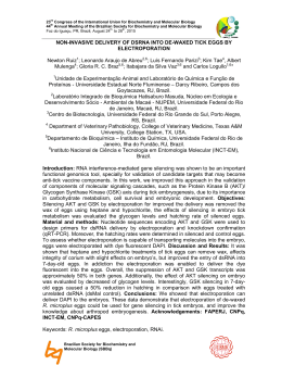
At figure 1 (A-F) are demonstrated the results of the interaction between P.
chlamydosporia isolates and A. suum eggs during the experiment.
All P. chlamydosporia isolates (Isol.5, Isol.31, Isol.1, VC1, Isol.12, Isol.22 and
VC4) showed the three types of effects (1, 2 and 3) at 5, 7, 10, 14, 15 and 21 days, but
there was not variation in ovicidal capacity (type 3 effect) among the isolates (P> 0.05)
throughout the experiment.
The following percentage results for type 3 effect were found for the isolates:
isolate 5 (35.0%, 25.0%, 52%, 28.32%, 52.34% and 58.33%), isolate 31 (6.68% 21.69%
60.89% 38.32% 46.34% and 58.32%), isolate 1 (31.67%, 30.0%, 36.68%, 35.0%, 32.16%
8
and 32.17%), isolate VC1 (31.69%, 48.83%, 45.9%, 26.68%, 56.67% and 57.67%), isolate
12 (26.67%, 23.33%, 34.2%, 26.0%, 30.0%, 48.0%), isolate 22 (11.66%, 33.34%, 25.0%,
33.33%, 39.83% and 53.17%) and isolate VC4 (41.67% 36.68% 36.67% 6.68% 48.33%
and 55.33%), at 5, 7, 10, 14, 15 and 21 days, respectively.
Light microscopy showed vegetative structures (hyphae) from P. chlamydosporia
colonizing eggs of A. sum. The subsequent egg rupture characterizes effect type 3.
Chlamydospore production by P. chlamydosporia was also observed in the Petri dishes
treated with the isolates.
A
B
Effect type 3
Effect type 1
Ascaris suum eggs
80
60
40
*
60
40
20
O
L
4
C
2
R
V
L.
2
L.
1
NT
O
IS
O
IS
O
C
treatment 5 days
2
1
V
L
C
1
1
IS
O
IS
O
L.
5
R
O
L
V
C
4
*
0
C
O
NT
22
12
IS
O
L.
IS
O
L.
V
C
1
L
1
31
IS
O
IS
O
L.
5
0
Effect type 3
80
L.
3
20
Effect type 2
100
IS
O
Effect type 2
100
IS
O
L.
Ascaris suum eggs
Effect type 1
treatment 7 days
*
C
D
Effect type 3
*
F
9
L
4
NT
RO
CO
VC
L.
22
IS
O
L.
12
IS
O
1
VC
1
IS
O
L
L.
31
0
IS
O
RO
L
4
20
NT
VC
40
treatment 14 days
E
60
O
treatment 10 days
Effect type 3
80
C
2
L.
2
IS
O
L.
1
2
1
IS
O
VC
L
IS
O
L.
L.
3
IS
O
IS
O
1
1
*
Effect type 2
100
IS
O
L.
5
40
20
0
Effect type 1
Ascaris suum eggs
Effect type 2
100
80
60
5
Ascaris suum eggs
Effect type 1
Effect type 3
60
40
*
20
80
60
40
20
*
4
VC
treatment 21 days
CO
IS
O
L.
22
1
VC
IS
O
L.
12
1
L
IS
O
L.
5
IS
O
L.
31
L
0
IS
O
VC
CO
treatment 15 days
Effect type 3
100
NT
RO
4
L.
22
IS
O
1
L.
12
IS
O
VC
1
IS
O
L
IS
O
L.
31
0
Effect type 2
Fig.1(A-F) - Percentages and standard deviation (bar) for the effects at type 1
3
+++
NT
RO
L
80
Effect type 1
Ascaris suum eggs
Effect type 2
100
IS
O
L.
5
Ascaris suum eggs
Effect type 1
+
,2
++
and
levels of the nematophagous fungus Pochonia chlamydosporia isolates (ISOL.5,
ISOL.31, ISOL.1, VC1, ISOL.12, ISOL.22 and VC4) compared with the control group
(without fungi) on Ascaris suum eggs at 5, 7, 10, 14, 15 and 21 interaction days.
+
Physiological and biochemical effects without morphological damage to the eggshell, with
hyphae adhered to the shell.
++
Lytic effects with morphological alterations of the embryo
and eggshell, without hyphal penetration.
+++
Lytic effect with morphological alteration of
the embryo and eggshell, in addition to hyphal penetration and internal colonization. *All
effects for all isolates significantly different from the controls (P<0.05).
4. Discussion
A fungus must be capable of internal egg colonization and destruction to be
considered ovicidal (Lysek et al. 1982). In the present work, all P. chlamydosporia isolates
showed type 3 effect, with hyphae penetrating A. suum eggs and destroying them,
characterizing the ovicidal activity.
P. chlamydosporia has proven action against eggs of parasitic nematodes of
animals and plants (Lopes et al. 2007; Araújo et al. 2008). Recent studies, under laboratory
conditions, have shown that it can also destroy eggs of trematodes, cestodes and nematodes
parasitizing domestic animals (Braga et al. 2007, 2008a, c; Araújo et al. 2008).
Isolates VC1 and VC4 were effective against A. suum eggs after 21 days of
incubation, with percentage results of 56.67% and 55.33%, respectively. Braga et al.
(2007) demonstrated the effectiveness of the P. chlamydosporia isolates VC1 and VC4
against eggs of Ascaris lumbricoides, a human gastrointestinal nematode, in intervals of 7,
10 and 14 days. At the end of the experiment they reported ovicidal percentages above
26% for both isolates.
10
Araújo et al. (2008) evaluated the effects of P. chlamydosporia isolates in
laboratory conditions and reported ovicidal activity (p <0.01) of VC1 and VC4 on A. suum
eggs with percentages of 13.0% and 17.3%, 13.9% and 17.7% and 19.0% and 20.0% at 7,
14 and 21 days respectively. In the present work, the isolates VC1 (48.83%, 26.68%,
56.67%) and VC4 (36.68%, 6.68%, 55.33%) showed better results for the same time
intervals. The other fungal isolates (Isol. 5, 31, 1, 12 and 22) were included to evaluate the
variability.
Braga et al. (2010) reported percentages of (31.5%, 39.4%) and (35.3%, 57.9%) for
type 3 effect of P. chlamydosporia (isolates 5 and 12) on Toxocara vitulorum, a bovine
gastrointestinal nematode, in laboratory conditions at intervals of 10 and 15 days
respectively. In this work, the same isolates were also effective against A. suum eggs.
Isolate 5 showed higher percentages for effect type 3 (52.0%, 52.34%) in the same
intervals. Braga et al. (2008c) stated that the penetration mechanism of ovicidal fungi in
parasitized eggs is still not completely elucidated and differences in the ovicidal activity
have been reported. Some authors have demonstrated that the enzymatic activity is one of
the main mechanisms used by fungi for of attack and penetration through egg shells
(Araujo et al. 2009; Braga et al. 2010). This may explain the differences in the percentage
results for effect type 3 between the two studies.
The first mode of mechanical action mentioned for an ovicidal fungus is the
appressorium, which is the structure used to penetrate the helminth eggs (Lysek, 1978).
Mizobutsi et al. (2000) evaluated the effect of 64 fungal isolates on Meloidogyne javanica
eggs and found that P. chlamydosporia showed the highest ovicidal activity, confirming its
importance as an ovicidal fungus.
The tested isolates (Isolates 31, Isol.1 and Isol.22) also showed ovicidal activity,
this is first study that demonstrated their action against gastrointestinal nematode parasites
of domestic animals.
Several field works aimed at controlling agricultural pests have been carried out
and demonstrated that P. chlamydosporia is effective in reducing nematode populations
(Kerry, 2001; Tobin et al. 2008). However, there is a scarcity of studies on the use of this
fungus to control helminthiasis in domestic animals.
Hansen and Perry (1994) pointed out serious concerns with the continued use of
antihelminthics. Parasite resistance to one or many anithelminthics is now widespread,
particularly in domestic animals, leaving many countries without means of controlling
11
gastrointestinal nematode parasitism. Therefore, the biological control of gastrointestinal
nematodiosis using ovicidal nematophagous fungi can be an important tool combined with
the chemical control.
The results of this work demonstrate that P. chlamydosporia (Isol.5, Isol.31, Isol.1,
VC1, Isol.12, Isol.22 and VC4) could be a potential biological control agent against A.
suum.
12
References
Araujo, J.M., Araújo, J.V., Braga, F.R., Carvalho, R.O., Silva, A.R. and Campos, A.K.,
2009. Interaction and ovicidal activity of nematophagous fungus Pochonia chlamydosporia
on Taenia saginata eggs, Experimental Parasitology, 121, 338-341
Araújo, J.V., Braga, F.R., Araújo, J.M., Silva, A.R. and Tavela, A.O., 2008. In vitro
evaluation of the effect of the nematophagous fungi Duddingtonia flagrans,
Monacrosporium sinense and Pochonia chlamydosporia on Ascaris suum eggs,
Parasitology Research, 102, 787-790
Araújo, J.V., Santos, M.A. and Ferraz, S., 1995. Efeito ovicida de fungos nematófagos
sobre ovos embrionados de Toxocara canis, Arquivo Brasileiro de Medicina veterinária e
Zootecnia, 47, 37-42
Araújo, J.V., Mota, M.A. and Campos, A.K., 2004. Controle biológico de helmintos
parasitos de animais por fungos nematófagos, Revista Brasileira Parasitologia Veterinária,
13,165-170
Ayres, M., Ayres, J.R.M., Ayres, D.L. and Santos, A.S., 2003. Aplicações estatísticas nas
áreas de ciências biológicas, (CNPq, Brasília).
Braga, F.R., Araújo, J.V., Campos, A.K., Caravalho, R.O., Silva, A.R., Tavela, A.O. and
Maciel, A.S., 2007. Observação in vitro da ação dos isolados fúngicos Duddingtonia
flagrans, Monacrosporium thaumasium e Verticillium chlamydosporium sobre ovos de
Ascaris lumbricoides (Lineu, 1758), Revista da Sociedade Brasileira de Medicina tropical,
40, 356-358
Braga, F.R.; Araújo, J.V.; Campos, A.K.; Araujo, J.M.; Silva, A.R.; Carvalho, R.O. and
Tavela, A.O., 2008a. In vitro evaluation of the effect of the nematophagous fungi
Duddingtonia flagrans, Monacrosporium sinense and Pochonia chlamydosporia on
Fasciola hepatica eggs, World Journal of Microbiology Biotechnology, 24, 1559-1564
Braga, F.R., Araújo, J.V., Campos, A.K., Silva, A.R., Araujo, J.M., Carvalho, R.O.,
Negrão-Correa, D. and Pereira, C.A.J., 2008b. In vitro evaluation of the effect of the
nematophagous fungi Duddingtonia flagrans, Monacrosporium sinense and Pochonia
13
chlamydosporia on Shistosoma mansoni eggs, World Journal of Microbiology
Biotechnology, 24, 2713- 2716
Braga, F.R., Araújo, J.V., Araujo, J.M., Carvalho, R.O., Silva, A.R., Campos, A.K. and
Tavela, A.O., 2008c. In vitro evaluation of the effect of the nematophagous fungi
Duddingtonia flagrans, Monacrosporium sinense and Pochonia chlamydosporia on
Moniezia sp eggs, Journal of Helminthology, 10, 241-243
Braga, F.R., Ferreira, S.R., Araújo, J.V., Araujo, J.M., Carvalho, R.O., Silva, A.R.,
Campos, A.K. and Freitas, L.G., 2010. Predatory activity of Pochonia chlamydosporia
fungus on Toxocara (syn. Neoascaris) vitulorum eggs. Tropical Animal Health and
Production, 42, 309-314
Gupta, N., Khan, D. K. and Santra, S.C., 2009. Prevalence of helminth eggs on vegetables
grown in wastewater-irrigated areas of Titagarth, West Bengal, India. Food Control, 20,
942-945
Hansen, J. and Perry, B., 1994. The epidemiology, diagnosis and control of helminth
parasites of ruminants, (Laboratory Research Animal Diseases, Nairobi)
Hurnik, D. and Dohoo, I.R., 1995. The impact of roundworms on swine production,
Compendium on Continuing Education for the Practicing Veterinary, 17, 589–593
Kerry,
B.R.,
2001.
Exploitation
of
the
nematophagous
fungal
Verticillium
chlamydosporium Goddard for the biological control of root-knot nematodes (Meloidogyne
spp). In: T. M. Butt, C. Jackson and N. Magan (eds), Fungi as Biocontrol Agents, (CAB
International, Wallingford), 155-167
Lopes, E.A., Ferraz, S., Ferreira, P.A., Freistas, L.G., Dhingra, O., Gardiano, C.G. and
Carvalho, S.L., 2007. Potencial de Isolados de fungos nematófagos no controle de
Meloidogyne javanica, Nematologia Brasileira, 31, 78-83
Lysek, H., 1978. A scanning electron microscope study of the effects of an ovicidal fungus
on the eggs of Ascaris lumbricoides, Parasitology, 77,139–141
14
Lysek, H., Fassatiová, O., Pineda, N.C., Hernández, N. and Lorenzo., 1982. Ovicidal fungi
in soils of Cuba, Folia Parasitologica, 29, 265-270
Massara, C.L., Ferreira, R.S., Andrade, L.D., Guerra, H.L. and Carvalho, O.S., 2003.
Atividade de dertegentes e desinfetantes sobre a evolução dos ovos de Ascaris
lumbricoides, Cadernos Saúde Pública, 18, 335-340
Mizobutsi, E., Ferraz, S.and Ribeiro, R.C.F., 2000. Avaliação do parasitismo de diversos
isolados fúngicos em ovos de Heterodera glycines e Meloidogyne javanica, Nematologia
Brasileira, 24, 167-172
Roepstorff, A. and Nansen, P., 1994. Epidemiology and control of helminth infections in
pigs under intensive and nonintensive production systems, Veterinary Parasitology, 54, 6985
Roepstorff, A., Nilsson, O., Oksanen, A., Gjerde, B., Richter, S.H., Ortenberg
E., Christensson, D., Martinsson, K.B., Bartlett, P.C., Nansen, P., Eriksen, L., Helle,
O., Nikander, S. and Larsen K., 1998. Intestinal parasites in swine in the Nordic Counties:
prevalence and geographical distribution, Veterinary Parasitology, 76, 305-319
Tobin, J.D., Haydock, P.P., Hare, M.C., Woods, S.R. and Crump, D.H., 2008. Effect of the
fungus Pochonia chlamydosporia and fosthiazate on the multiplication rate of potato cyst
nematodes (Globodera pallida and G. rostochiensis) in potato crops grown under UK field
conditions, Biological Control, 46, 194-201
Urquhart, G.M., Armour, J., Duncan, J.L., Dunn, A.M. and Jennings. F.W., 1998.
Parasitologia Veterinária, (Guanabara & Koogan, Rio de Janeiro).
Wagner, B. and Polley, L., 1997. Ascaris suum prevalence and intensity: an abattoir survey
of market hogs in Saskatchewan, Veterinary Parasitology, 73, 309–313
15
Capítulo 2
Biological control of Ascaris suum eggs by Pochonia chlamydosporia fungus
Veterinary Research Communications, in press (impact factor 1.05)
DOI: 10.1007/s11259-011-9494-6
16
Abstract
Ascaris suum is a gastrointestinal nematode parasite of swines. The aim of this study was
to observe Pochonia chlamydosporia fungus on biological control of A. suum eggs after
fungus passage through swines gastrointestinal tract. Eighteen pigs, previously dewormed,
were randomly divided into three groups: group 1, treated with the fungus isolate VC4;
group 2, treated with the fungus isolate VC1 and group 3 did not receive fungus (control).
In the treated groups, each animal received a 9g single dose of mycelium mass containing
P. chlamydosporia (VC1 or VC4). Thereafter, animal fecal samples were collected at the
following intervals: 8, 12, 24, 36, 48, 72 and 96 hours after treatment beginning and these
were poured in Petri dishes containing 2% water-agar culture medium. Then, one thousand
A. suum eggs were poured into each dish and kept in an incubator at 26oC and in the dark
for 30 days. After this period, approximately one hundred eggs were removed from each
Petri dish and morphologically analyzed under light microscopy following the ovicidal
activity parameters. The higher percentage observed for isolated VC4 eggs destruction was
57.5% (36 hours) after fungus administration and for isolate VC1 this percentage was
45.8% (24 hours and 72 hours) (p>0.01). P. chlamydosporia remained viable after passing
through the gastrointestinal tract of swines, maintaining its ability of destroying A. suum
eggs.
Keywords: Ascaris suum, Pochonia chlamydosporia, Nematophagous fungi, Biological
control
Introduction
Swines are affected by many parasites species resulting in considerable production
losses, as a result of the low feed conversion rate and animal growth, increased
susceptibility to other diseases and organ condemnation at slaughter (Wagner and Polley
1999). Among the parasites, stand out the gastrointestinal helminths, represented by
nematodes of the genera: Ascaris, Oesophagostomum, Strongyloides, Trichuris and
17
Hyostrongylus prevalent in different geographic areas (Roepstorff et al. 1998; D'Alencar et
al. 2006).
Ascaris suum is common among swines raised in extensive management systems in
the world (Nansen and Roepstorff 1999). This parasite is usually associated with liver
damages ("milk spots" caused by larvae migration), resulting in organ condemnation. A.
suum eggs stand out for their resistance in the environment, remaining viable for long
periods (Urquhart et al. 1998). Like other ascarids, it eggs can contaminate soil, water and
food (Paulino et al. 2001; Gupta et al. 2009), once present in the food they are not easily
removed, as they have a great capacity of adhering to surfaces (Massara et al. 2003). The
changes in breeding system can decrease this nematode infection rates, however, the
infection agents can persist even in farms with good management practices (Roepstorff and
Nansen 1994).
Among the natural antagonists of gastrointestinal helminthes, for eggs and larvae
present in environment, nematophagous fungi stand out (Gronvold et al. 1996; Larsen
1999). Several works have reported the success of these fungi in biological control under
laboratory conditions (grown fungus on solid medium, in vitro) and in natural conditions,
passage through domestic animals gastrointestinal tract, with germination and growth in
feces (Braga et al. 2010a; Tavela et al. 2010). Among the fungi species used, P.
chlamydosporia has been successfully tested in vitro (Ferreira et al. 2010) and after passing
through the gastrointestinal tract of domestic animals (Braga et al. 2010a; Singh et al.
2010), however, there is no report about its passage through gastrointestinal tract of swines
with subsequent germination and growth.
The aim of this study was to observe P. chlamydosporia fungus on biological
control of A. suum eggs after fungus passage through swines gastrointestinal tract.
Materials and methods
Study area
The experiment was conducted at the Swine experimental sector of the Federal University
of Viçosa, located in the city of Viçosa, Minas Gerais, Brazil, latitude 20° 45'20 "and
longitude 42° 52'40.
Fungi strains
18
Two fungal isolates (VC4 and VC1) of P. chlamydosporia, originated from
Brazilian soil, were tested for their ability passing through gastrointestinal tract of swines.
Disc cultures of isolates (VC1 and VC4) kept in test tubes containing the culture medium
2% water-agar (2% WA) were transferred to Erlenmeyer flasks containing 150ml of liquid
medium GPY (glucose, sodium peptone and yeast extract), and these were kept shaking at
120 rpm in the dark at a temperature of 26oC for 10 days. After this period, the mycelia
were removed, filtered and weighed on analytical balance (Braga et al 2010).
Obtaining of A. suum eggs
The eggs of A. suum were recovered from the dissection of a mature female worm
obtained from a swine which died of natural causes. Then, these eggs were
morphologically analyzed under light microscopy, objective lens 10x, according to the
description of Urquhart et al. (1998). Thereafter, the eggs were washed by centrifugation
10 times in distilled water and centrifuged at 1,000 rpm for 5 minutes each time. The
supernatant was discarded at the end of each centrifugation cycle. Eggs were then
incubated to embryonate at 25ºC for 21 days with a solution containing 0.005%
streptomycin sulphate and 0.01% chloramphenicol as described by Araújo et al. (1995).
In vivo essay
It was used eighteen crossbred swines, male, with average weight of 32.08 kg (±
1.83), kept in masonry bays, previously dewormed with swines anthelmintic, febemdazole
4% (Intervet®) orally, using a dose of 0.12 mg per kilogram of body weight, 14 days
before receiving the fungal treatment, according to the methodology described by Braga et
al. (2010a). Then, the animals were divided into three groups, with 6 animals each. (1)
group treated with the fungus (VC4), (2) group treated with the fungus (VC1) and (3)
group that did not receive fungus (control). In the treated groups (1 and 2) each animal
received 9g of mycelium fungi mass in a single dose, containing P. chlamydosporia
isolates (VC4 or VC1) mixed in 100 grams of swine commercial food (cottonseed meal,
sodium chloride, Corn gluten meal 21, soybean meal, wheat bran, flour of meat and bones
of cattle, dicalcium phosphate, corn germ, ground whole corn, Mineral premix and vitamin
premix) according to the methodology described by Braga et al. (2010a).
Fecal samples from each animal were collected at 8, 12, 24, 36, 48, 72 and 96 hours
interval after the fungus administration. Samples were homogenized; and feces (2 g) were
19
removed of each sample and placed in Petri dishes, 9 cm diameter, containing 2% WA, for
each interval collected there was 18 dishes, six dishes for each group. One thousand
embryonated eggs of A. suum were placed in each dish of treated and control groups, and
incubated at 26° C in the dark for 30 days (Braga et al 2010). Petri dishes of treated and
control groups were examined daily to assess P. chlamydosporia (VC1 or VC4)
characteristic structures, such as conidia, conidiophores and chlamydospores, which were
analyzed according to the classification proposed by Zare et al. (2001).
After thirty days, one hundred eggs were removed from each Petri dish of treated
and control groups according to the technique described by Araújo et al. (1995) and placed
on glass slides with a drop of Aman blue 1%. Eggs were examined under a light
microscope (40X objective lens) using the parameters established by Lysek et al. (1976):
effect of type 1; type 2 and type 3 (eggs destruction).
Statistical analysis
Data were submitted to Friedman at 1% probability non parametric statistical test (Ayres et
al. 2007).
Results
At Table 1 are demonstrated the results of P.chlamydosporia, isolates VC4 or VC1,
ovicidal activity on Ascaris suum eggs in different intervals (8, 12, 24, 36, 48, 72 and 96
hours). The first characteristic structures of P.chlamydosporia to be observed in dishes of
treated groups with VC1 and VC4 were the chlamydospores. Later, during the study, it was
also possible to verify P. chlamydosporia typical hyphae and conidia presence on the
dishes of the treated groups. P. chlamydosporia showed ovicidal activity against A. suum
eggs after passing through swines gastrointestinal tract, and this activity remained at all
intervals. In the dishes of the control group (without fungus) was not observed the presence
of chlamydospores, hyphae and conidia of P. chlamydosporia.
Although isolate VC4 showed a higher percentage of type 3 effect, observed most
of the time, there was not statistical difference between the isolates tested (p >0.01)
referring to ovicidal activity.
20
Table 1-Percentages and standard desviations of ovicidal activity of the isolates (VC1 and VC4) Pochonia
chlamydosporia and control against Ascaris suum eggs referring to the collection of 8, 12, 24, 36, 48, 72
and 96 hours, after 30 days of interaction. Groups
VC4
VC1
Control
effect type 1*
49.8A8.1
47.5 A 8.8
0 B 0
VC4
VC1
Control
31.6 A 11.2
30.0 A 15.1
0 B 0
VC4
VC1
Control
15.0 A 6.3
16.6 A 8.7
0 B 0
VC4
VC1
Control
15.0 A 4.4
19.1 A 8.0
0 B 0
VC4
VC1
Control
18.3 A 10.3
38.1 A 5.2
0 B 0
VC4
VC1
Control
15.0 A 8.3
19.2 A 8.0
0 B 0
VC4
VC1
Control
18.3 A 8.7
22.5 A 11.2
0 B 0
Effect at 8 hours
effect type 2**
29.3 A 2.1
27.5 A 6.8
0 B 0
Effect at 12 hours
33.3 A 9.8
33.3 A 8.7
0 B 0
Effect at 24 hours
32.5 A 6.1
38.3 A 6.8
0 B 0
Effect at 36 hours
27.5 A 7.5
30.8 A 8.1
0 B 0
Effect at 48 hours
31.6 A 5.1
32.1 A 5.2
0 B 0
Effect at 72 hours
28.3 A 5.1
35.0 A 6.3
0 B 0
Effect at 96 hours
40.8 A 6.6
32.5 A 4.1
0 B 0
effect type 3***
20.8 A 6.8
26.6 A 4.0
0 B 0
35.0 A 10.9
36.6 A 10.3
0 B 0
52.5 A 6.1
45.8 A 9.9
0 B 0
57.5 A 11.2
50.0 A 6.3
0 B 0
50.0 A 8.3
29.6 A 1.8
0 B 0
56.6 A 7.5
45.8 A 4.9
0 B 0
40.8 A 5.8
45.0 A 8.9
0 B 0
Percentages followed by the same letter, in the line, are not significantly different (p>0.01). *Physiological and
biochemical effects without morphological damage to the eggshell, with hyphae adhered to the shell. **Lytic effects
with morphological alterations of the embryo and eggshell, without hyphal penetration. ****Lytic effect with
morphological alteration of the embryo and eggshell, in addition to hyphal penetration and internal colonization.
21
Discussion
Fungus utilization in intestinal parasites helminths eggs and larvae biological
control must pass through domestic animals gastrointestinal tract, grow in feces and
predate nematode eggs (Larsen, 1999). One of the factors that can favor this passage is
chlamydospores production (resistance structures). In the present work, chlamydospores
production in Petri dishes of groups treated with P. chlamydosporia (VC1 and VC4) was
observed, demonstrating that the fungus might be able to survive after passing through
swines gastrointestinal tract. Still, for chlamydospores production is important to stand out
viability this structure for its use in environmental biological control (Larsen 1999; Terril
et al. 2004)
The present study showed A. suum eggs destruction (type 3 effect) by P.
chlamydosporia isolates (VC4 and VC1) after a long period of interaction. These results
are in agreement with Ferreira et al. (2010) and Araújo et al. (2008), that also reported the
destruction of A. suum eggs after twenty one days of interaction in vitro,
with P.
chlamydosporia (VC4 and VC1) grown in 2%WA medium. However, these studies have
been conducted only in laboratory conditions, the fungus was already grown. In this study
the fungus P. chlamydosporia passed through the swines gastrointestinal tract, germinated
from their feces and destroyed the A. suum eggs at the end of thirty days of interaction.
Still in comparisons to the work of Ferreira et al. (2011) and Araújo et al. (2008), the
obtained destruction percentages for VC4 and VC1 were the following: (57.67% and
55.3%) and (19% and 20%), respectively, at the end of study. The results in the present
study were similar (Table 1), at little different conditions. Costa et al. (2001) argued that
2%WA medium did not reflect the environmental conditions and so little the habitat in
which nematophagous fungi (natural antagonists) are present. In that way, studies that
might mimic these fungi environment, in this case the fecal environment, are important.
Moreover, according to Araújo et al. (2010) predatory and ovicidal nematophagous fungi
are generally saprophytic and therefore should be routinely tested under experimental
conditions in order to prove its predatory activity in adverse conditions.
Present results obtained can be compared to what is already known in the literature,
being that: Lysek (1976) reported that fungi may be considered as ovicidal if it presents
type 3 effect-destruction of eggs. De et al. (2008) and Braga et al. (2010a) investigated P.
chlamydosporia ovicidal activity against trematode and nematode eggs and noted that this
22
fungus was effective destructing eggs. The present study is in accordance with the
literatures.
According to Braga et al. (2010b) it is important that the fungus have quick ovicidal
action and that it remains for long time, thus, it can be to used in control of helminth eggs
with short and long hatching period. The results presented and discussed in this study
suggest that P. chlamydosporia could be used in biological control of A. suum eggs present
in the environment.
References
Araújo JM, Braga FB, Araújo JV, Soares FEF, Geniêr HLA (2010) Biological control of
Taenia saginata eggs. Helminth 47:189 – 192. DOI: 10.2478/s11687-010-0028-5.
Araújo JV, Braga FR, Araujo JM, Silva AR, Tavela AO (2008) In vitro evaluation of the
effect of the nematophagous fungi Duddingtonia flagrans, Monacrosporium sinense and
Pochonia chlamydosporia on Ascaris suum eggs. Parasitol Res 102:787-790. DOI:
10.1007/s00436-007-0852-9.
Araújo JV, Santos MA, Ferraz S (1995) Efeito ovicida de fungos nematófagos sobre ovos
embrionados de Toxocara canis. Arq. Bras Med Vet Zootec 47:37- 42.
Ayres M, Ayres JRM, Ayres DL, Santos AS (Eds.) (2007): Aplicações estatísticas nas
áreas das ciências biológicas e médicas. Sociedade Civil Mamirauá . 364p.
Braga FR, Araújo JV, Silva AR, Carvalho RO, Araujo JM, Ferreira SR, Carvalho GR
(2010a) Viability of the nematophagous fungus Pochonia chlamydosporia after passage
through
the
gastrointestinal
tract
of
horses.
Vet
Parasitol
168:264-268.
DOI:10.1016/j.vetpar.2009.11.020.
Braga FR, Araújo JV, Carvalho RO, Silva AR, Araujo JM, Freitas FES, Geniêr HLA,
Ferreira SR, Queiroz JH (2010b) Ovicidal action of a crude enzymatic extract of the
fungus Pochonia chlamydosporia against cyathostomin eggs. Vet Parasitol 172:264-268.
DOI:10.1016/j.vetpar.2010.05.011.
Costa MJN, Campos VP, Pfenning LH, Oliveira DF (2001) Toxicidade de filtrados
fúngicos a Meloidogyne Incognita. Fitopatol. Bras. 26:749-755.
23
D’Alencar AS, Faustino MAG, Sousa DP, Lima MM, Alves LC (2006) Infecção por
helmintos e coccídios em criação de suínos de sistema confinado localizada no município
de Camaragibe-Pe. Ciência Vet Tróp 9:79 – 86.
De S, Sanyal PK, Sarkar AK, Patel NK, Pal S, Mandal SC (2008) Screening for Indian
isolates of egg-parasitic fungi for use in biological control of fascioliasis and
amphistomiasis
in
ruminant
livestock.
J
Helminthol
82:271–277.
DOI:
10.1017/S0022149X08982602.
Ferreira SR, Araújo JV, Braga FR, Araujo JM, Carvalho RO, Silva AR, Frassy LN, Freitas
LG (2010) Ovicidal activity of seven Pochonia chlamydosporia fungal isolates on Ascaris
suum eggs. Trop. Animal Health and Prod 43:639-642. DOI: 10.1007/s11250-010-9744-6.
Gronvold J, Henriksen SA, Larsen M, Nansen P, Wolstrup J (1996) Biological control
Aspects of biological control ,with special reference to arthropods, protozoans and
helminths of domesticated animals. Vet Parasitol 64:47-64. DOI:10.1016/03044017(96)00967-3.
Gupta N, Khan DK, Santra SC (2009) Prevalence of helminth eggs in vegetables grown in
wastewater-irrigated areas of Titagarth, West Bengal, India. Food Control 20:942-945.
DOI:10.1016/j.foodcont.2009.02.003.
Larsen M (1999) Biological control of helminths. Int J Parasitol 29, 139– 146.
DOI:10.1016/S0020-7519(98)00185-4
Lysek, H., 1976. Classification of ovicide fungi according to type of ovicidity. Acta Univ.
Palack. Olomue 76:9–13.
Massara CL, Ferreira RS, Andrade LD, Guerra HL, Carvalho OS (2003) Atividade de
detergentes e desinfetantes sobre a evolução dos ovos de Ascaris lumbricoides. Cadernos
Saúde Pub 18:335-340.
Nansen P, Roepstorff A (1999) Parasitic helminths of the pig: factors influencing
transmission
and infection levels. Inter J Parasitol 29:877-891. DOI:10.1016/S0020-
7519(99)00048-X.
24
Paulino RC, Castro EA, Soccol VT (2001) Tratamento anaeróbico de esgoto na redução da
viabilidade de ovos de helmintos. Rev Soc Bras Med Trop 34:421-428
Roepstorff A, Nilssonb O, Oksanenc A,
Gjerded B, Richtere SH, Ortenbergf E,
Christenssonf D, Martinsson KB, Bartletth PC, Nansena P, Eriksena L, Helled O,
Nikanderc S, Larseni K (1998) Intestinal parasites in swine in the Nordic Countries:
prevalence and geographical distribution. Parasitol 76:305-319. DOI:10.1016/S03044017(97)00223-9.
Roepstorff A, Nansen P (1994) Epidemiology and control of helminth infections in pigs
under intensive and non-intensive production systems. Vet Parasitol 54:69–85.
DOI:10.1016/0304-4017(94)90084-1
Singh RK, Sanyal PK, Patel NK, Sarkar AK, Santra AK, Pal1 S, Mandal SC (2010)
Fungus–benzimidazole interactions: a prerequisite to deploying egg-parasitic fungi
Paecilomyces lilacinus and Verticillium chlamydosporium as biocontrol agents against
fascioliasis and amphistomiasis in ruminant livestock. J Helminthol 84:123–131. DOI:
10.1017/S0022149X0999034.
Tavela AO, Araújo JV, Braga FR, Silva AR, Araujo JM, Carvalho RO, Ferreira SR,
Carvalho GR (2010) Biological control of cyathostomin (Nematoda: Cythostominae) with
nematophagous fungus Monacrosporium thaumasium tropical southeastern Brazil. Vet
Parasitol 175:92-96. DOI:10.1016/j.vetpar.2010.09.035
Terrill TH, Larsen M, Samples O, Husted S, Miller JE, Kaplan RM, Gelaye S (2004)
Capability of the nematode-trapping fungus Duddingtonia flagrans to reduce infective
larvae of gastrointestinal nematodes in goat faeces in the southeastern United States: dose
titration
and
dose
time
interval
studies.
Vet
Parasitol
120:285–296.
DOI:10.1016/j.vetpar.2003.09.024.
Urquhart GM, Armour J, Duncan JL, Dunn AM, Jennings FW (1998) Parasitologia
Veterinária. Guanabara Koogan, Rio de Janeiro, pp. 373.
Wagner B, Polley L (1999) Ascaris suum: seasonal egg development rates in a
Saskatchewan
pig
barn
Veterinary
Parasitology
4017(99)00102-8.
25
85:71–78.
DOI:10.1016/S0304-
Zare R, Gams W, Evans HC (2001) A revision of Verticillium section Prostrata V. The
genus Pochonia, with notes on Rotiferophthora. Nova Hedwigia 73:51-86
26
Capítulo 3
Nematophagous fungi against infective larvae Oesophagostomum spp of pigs
Article submitted to Veterinarni Medicina (impact factor 0.644)
27
Abstract
Pigs are affected by many species of gastrointestinal nematodes of the genera Ascaris and
Oesophagostomum. Two experimental assays (A and B) evaluated the action of conidia of
the nematophagous fungi Duddingtonia flagrans (AC001), Monacrosporium sinense
(SF53) and Artrhobotrys robusta (I-31) against infective larvae (L3) of Oesophagostomum
spp in 2% water-agar (2%WA) medium and coprocultures. The first assay consisted of
three groups of 1000 Oesophagostomum L3 treated with 1000 conidia of isolates AC001,
SF53 and I-31 and control without fungus plated in 2% WA. In the second assay, 1000
conidia of the same isolates were added to 20g of feces and incubated at 26°C for 8 days.
The L3 were recovered after this period. Fungal treatments in the first assay were efficient
in the capture of Oesophagostomum spp L3 (p<0.01) when compared to the control
treatment(without fungi). There was no variation in the predatory capacity among the
tested fungal isolates (p>0.01) during the experimental period of seven days. There were
significant reductions (p<0.01) of 94.4%, 92.9% , and 88.3% in the means of
Oesophagostomum L3 recovered from the treatments with isolates AC001, SF53 and I-31,
respectively, when compared to the control treatment. Significant difference (p<0.01) was
found between the number of L3 recovered from the treated groups and the control group at
the end of assay A. Assay B also showed statistical differences (p<0.01) between the
means of recovered L3 in the treated groups and the control: 75.3% (AC001), 63.7%
(SF53) 63.3% (I-31). In this study, the three isolates of predatory fungi D. flagrans
(AC001), M. sinense (SF53) and A. robusta (I-31) were efficient in the in vitro capture and
destruction of Oesophagostomum L3 and are potential biological control agents of this
nematode.
Keywords: Biological control, nematophagous fungi, Oesophagostomum, pigs.
1. Introduction
Helminths that infect pigs are widely varied in size, life cycle and pathogenicity.
Diseases caused by internal and external parasites are reported in pig farms
worldwide. The infections are not always apparent and persist at subclinical levels for
extended periods, leading to death of animals (Sobestiansk et al., 1998). The genus
Oesophagostomum, with worldwide distribution, it is among the main intestinal helminths
28
infecting pigs. The major species are O. dentatum and O. quadrispinulatum (Stewart and
Gasbarre, 1989). Oesophagostomum infection occurs through the ingestion of infective
larvae (L3) present in the environment. Roepstorff and Jorsal (1989) reported the
occurrence of helminths in 66 pig herds in Denmark, with 58% of Oesophagostomum spp.
Roepstorff and Nansen (1994) pointed out that the expanding technology in
production systems improves sanitary conditions by creating monitoring systems that
reduce chances of parasite transmission. However, pigs raised extensively on pasture
demand reformulated control measures against parasites, with special attention to the
control of free-living infective stages. In this case, the control with nematophagous fungi is
suggested because their action is concentrated in the fecal environment and directed
against the free-living forms (Larsen, 1999).
Nematophagous
fungi
are
classified
into
predators,
opportunists
and
endoparasites. The genera Duddingtonia, Monacrosporium and Arthrobotrys of the
predator group are the most studied for the control of nematodiosis in domestic animals
(Braga et al., 2010a). D. flagrans is considered the most promising because of the
production of large amounts of chlamydospores. M. thaumasium and A. robusta have been
successfully used in the control of infective larvae of gastrointestinal nematodes of
domestic animals in laboratory and field conditions (Carvalho et al., 2010; Silva et al.,
2010; Silva et al., 2010). However, there are not reports on the action of nematophagous
fungi in the control of gastrointestinal nematodes of pigs.
This study evaluated the in vitro predatory activity of the fungi D. flagrans,
M. sinense and A. robusta against larvae of Oesophagostomum spp in two experimental
assays.
2. Material and Methods
2.1. Fungal culture
Isolates of D. flagrans (AC001), M. sinense (SF53) and A. robusta (I-31) were
obtained from a Brazilian soil, in Viçosa, Zona da Mata region, State of Minas Gerais,
between 20 45'20"S and 42 52'40" W longitude, 649 m altitude. The isolates were kept
in test tubes with 2% corn-meal-agar (2% CMA), at 4°C in the laboratory of Parasitology
of the Federal Universidad of Viçosa.
2.2. Conidia Collection
29
Culture disks (4 mm in diameter) of fungal isolates in 2% CMA were removed
from the test tubes and transferred to 9.0 cm Petri dishes, containing 20 ml of 2% potato
dextrose agar and kept at 25 °C in the dark for 10 days. After growth, new culture disks (4
mm in diameter) were transferred to 9.0 cm diameter Petri dishes containing 20 ml of 2%
water-agar (2% WA). Distilled water (1 ml) containing 1000 larvae of Panagrellus sp was
daily added to the plates to induce conidial formation for 21 days. After complete fungal
development, 5 mL of distilled water were added to each plate and conidia and mycelial
fragments were removed using technique described by Carvalho et al. (2009).
2.3. Oesophagostomum spp L3
Infective larvae (L3) of Oesophagostomum spp. were recovered from feces of
naturally infected pigs using vermiculite coproculture for 15 days. At the end of this
period, third stage larvae (L3) were obtained by the Baermann method, with water at 4245oC and 12h decantation time. Larvae were identified according to Ueno and Gonçalves
(1998).
2.4. Assays
Two in vitro assays (A and B) were carried out with an interval of eight
days. Assay A evaluated fungal activity of conidia of D. flagrans (AC001), M. sinense
(SF53) and A. robusta (I-31) on Oesophagostomum L3 in 2% WA. Assay B evaluated the
action of conidia of isolates AC001, SF53 and I-31 in coprocultures containing
Oesophagostomum larvae.
2.5. Assay A
Assay A consisted of four treatments of fungal isolates (AC001, SF53 and I-31) and
a control without fungus plated in 9.0cm Petri dishes containing 20 ml of 2%WA, with six
repetitions each. Petri dishes were marked into 4mm fields. A thousand Oesophagostomum
L3 were plated with 1000 conidia of the fungal isolates. The control contained 1000 L3
without fungus, according to the methodology used by Braga et al. (2010a). Ten random
fields (4mm diameter) were examined per plate of the treated and control groups, using an
optical microscope (10 X objectives) for L3 counts, every 24 hours, for seven days. After 7
30
days, the non-predated L3 were recovered from the Petri dishes using the Baermann
method, with water at 42 ºC (Araújo et al., 1993).
2.6. Assay B
Fresh feces with positive EPG were used for preparing coprocultures, which were
mixed with autoclaved and moistened vermiculite. Treatments consisted of four groups of
fungal isolates (AC001, SF53 and I-31) and a control without fungus, with six repetitions
each. Each replicate received 1000 fungal conidia. The control group contained fecal
culture only. Coprocultures, from treated and control groups, were incubated at 26 °C and
for 8 days. At the end of this period, L3 were recovered by the Baermann method, which
were identified and quantified according to the criteria described by Ueno and Gonçalves
(1988) under optical microscope (10 x objectives).
2.7. Statistical analysis
Means of Oesophagostomum L3 recovered from tests A and B were examined by
analysis of variance at 1% probability level (Ayres et al. 2007). Predation efficiency of L3
relative to the control group was assessed by the Tukey’s test at 1% probability level. The
percent reduction in means of recovered L3 was calculated by the following equation:
Reduction % = (Mean of L3 recovered from control group - Mean of L3 recovered from treated group) x 100
Mean L3 recovered from control group
3. Results and Discussion
Isolates of predatory fungi D. flagrans (AC001), M. sinense (SF53) and A. robusta
(I-31) were capable to prey Oesophagostomum L3 in assay A.
No significant difference (p>0.01) was found in the comparison of capture and
destruction of Oesophagostomum L3 among plates of the groups treated with isolates
AC001, SF53 and I-31 during the experimental assay (Table 1). However, there was
difference (p<0.01) between the means of non-predated Oesophagostomum L3 per 4mm
diameter field in the plates of the control group and means of L3 recorded in the fungitreated groups. The recorded percent reduction in Oesophagostomum L3 were: 94.4%
(AC001), 92.9% (SF53) and 88.3% (I-31).
31
Table 1- Daily means and standard deviation (±) of infective larvae (L3) not predated of
Oesophagostomum spp by field 4 mm diameter in 2% water-agar during the period of seven days,
for treatments with the fungal isolates
Duddingtonia flagrans (AC001), Monacrosporium
sinense (SF53), Arthrobotrys robusta (I-31) and in control without fungus.
Treatments
Time
(Means of non-predated Oesophagostomum L3 per 4mm)
(days)
AC001
SF53
I31
Control
1
3.86a b ± 2.4
4.98a ± 2.8
1.78b ± 1.0
11.8c ± 7.6
2
1.21a ± 1.26
1.41a ± 1.02
1.43a ± 0.72
4.15b ± 5.15
3
1.36a ± 1.17
1.76a ± 1.35
1.5a ± 1.2
2.53b ± 1.56
4
1.4a ± 1.2
1.8a ± 1.3
1.4a ± 1.3
2.5b ± 1.5
5
1.5a ± 1.08
1.68a ± 0.9
1.18a ±0.8
5.01b ± 4.6
6
0.73a ± 0.8
0.71a ± 0.9
0.91a ± 0.9
4.81b ± 3.6
7
1.08a ± 1.0
1.8a ± 1.4
2.96a ± 2.7
11.98b ± 7.7
Means followed by at least one common letter, in the lines, are not significantly different by the Tukey’s test at a
1% probability level.
Nematophagous fungi were not observed in plates of the control group during the
experiment, so this shown variation in the percentage of larvae observed in the table,
probably, it is due to migration of larvae to regions of the plates where there was more
humidity. The presence of Oesophagostomum L3 was essential for trap formation in plates
containing nutrient-poor medium such as 2% WA. Evidence of predation was confirmed
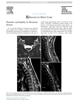
by the means of recovered L3 using the Baermann method 7 days post-plating, at the end of
the assay A (Fig. 1). Difference (p<0.01) was found between the number of recovered L3 in
the treated groups and the control without fungi.
32
*
Fig. 1 -Means and standard deviation (bars) of infective non-predated Oesophagostomum
spp. larvae recovered from 2% water-agar plates by the Baermann method on the seventh
day of treatment with the following fungal isolates: Duddingtonia flagrans (AC001),
Monacrosporium sinense (SF53), Arthrobotrys robusta (I-31) and a control group (without
fungi). Asterisk denotes significant difference (p<0.01) between the fungus-treated group
and the control - Tukey’s test at a 1% probability level.
At the end of 8 days, means of recovered larvae in assay B showed that fungal
conidia of D. flagrans (AC001), M. sinense (SF53) and A. robusta (I-31) were effective in
reducing Oesophagostomum L3 (Fig. 2). Significant difference (p<0.01) was found
between the means of larvae recovered from the treated groups and the control group with
the following percent reductions: 75.3% (AC001), 63.7% (SF53) and 63.3% (I-31).
33
*
Fig. 2 - Means and standard deviation (bars) of infective non-predated Oesophagostomum
spp. larvae recovered from coproculture by the Baermann method on the seventh day of
treatment with the following fungal isolates: Duddingtonia flagrans (AC001),
Monacrosporium sinense (SF53), Arthrobotrys robusta (I-31) and a control group (without
fungi). Asterisk denotes significant difference (p<0.01) between the fungus-treated group
and the control - Tukey’s test at a 1% probability level.
The present study showed that conidia of predatory fungi D. flagrans, M. sinense
and A. robusta were capable of preying (p>0.01) Oesophagostomum spp. L3 at the end of
the experimental assay A. Isolate AC001 showed efficiency in the capture and destruction
of L3, with percent reduction of 94.4% L3 recovered from the plates after 7 days. These
results are consistent with reports by Braga et al. (2010a) that isolate AC001 was more
efficient in preying Ancylostoma ceylanicum L3, a geohelminth that affects dogs. Braga et
al. (2010b) also showed that isolate AC001 stored in silica-gel for seven years was
efficient in the capture and destruction of Haemonchus contortus L3. These findings
confirm the efficiency and applicability of D. flagrans (AC001).
M. sinense (SF53) and A. robusta (I-31) were also shown efficient in the capture
and destruction of Oesophagostomum spp L3 with percent reductions of 92.9% and 88.3%,
7 days post-plating. Araujo et al. (2010) reported recently that M. thaumasium (NF34) and
A. robusta (I-31) effectively captured and destroyed Strongyloides westeri L3. These
34
results show that the nematophagous fungi can be used to control a wide range of larvalstage nematodes.
According to Nansen et al. (1988), a greater mobility of nematodes increases the
stimulus for trap-formation and predation of nematophagous fungi. This was also found in
our study, as we observed predation of L3 from the first day, after 24 hours of interaction.
In assay B, conidia of isolates AC001, SF53 and I-31 were effective in reducing
(p>0.01) Oesophagostomum L3 of coprocultures. However, at 8 days post-plating, isolate
AC001 showed the highest percent reduction (75.3%). In a recent study, Silva et al. (2010)
reported that in the concentration of 1000 conidia, AC001 was effective in reducing
H. contortus L3 after eight days of coproculture, providing for an 87.5% percent reduction.
Braga et al. (2009) showed that AC001 was effective in reducing cyathostomin L3
recovered from coprocultures of horses with positive EPG. In the present study, conidia of
isolates M. sinense (63.7%) and A. robusta (63.3%) in the same concentrations were also
effective in reducing the Oesophagostomum L3 in the same interval. Araújo et al. (2006)
found no difference (p>0.05) between the associations and the various fungal isolates,
including M. sinense and A. robusta, used to prey Cooperia sp. and Oesophagostomum sp.,
which agrees with our findings, indicating that nematophagous fungi can be successfully
used to reduce recurrent infections.
The difference between percent reduction of larvae of the tests A and B, (AC001),
92.9% (SF53), 88.3% (I-31) and 75.3% (AC001), 63.7% (SF53), 63.3% (I-31),
respectively, it is probably related to the fact that the environment of the coprocultures
there is greater amount of organic matter and as fungi are saprophyte, they had part of ther
action was not directed to the larvae but to for organic matter.
Choosing a nematophagous fungus that produces effective results in nematode
control should consider both in vitro and field tests. Our results suggest the need for future
field studies using AC001, SF53 and I-31 against gastrointestinal nematodes in pigs.
Here, we report that conidia of D. flagrans (AC001), M. sinense (SF53) and
A. robusta (I-31) were efficient in the capture and destruction of L3 of Oesophagostomum
spp. The fungi caused reduction in the number of L3 recovered from the coprocultures. The
main route of infection of Oesophagostomum spp. is the ingestion of infective L3 present in
the environment. Thus, the findings of this study show that these fungi can help in the
control of this nematode.
35
References
Araujo JM, Araújo JV, Braga FR, Carvalho RO (2010): In vitro predatory activity of
nematophagous fungi and after passing through gastrointestinal tract of equine on infective
larvae of Strongyloides westeri. Parasitology Research 107, 103-108.
Araujo JV, Assis RCL, Campos AK, Mota MA (2006): Antagonistic effect of
nematophagous fungi Monacrosporium, Arthrobotrys and Duddingtonia on infective
Cooperia sp. and Oesophagostomum sp. Larvae (In Portuguese) . Arquivo Brasileiro de
Medicina Veterinária e Zootecnia 58, 373-380.
Araújo JV, Santos MA, Ferraz S, Maia AS (1993): Antagonistic effect of predacious
Arthrobotrys fungi on infective Haemonchus placei larvae. Journal of Helminthology 67,
136–138.
Ayres M, Ayres JRM, Ayres DL, Santos AS (Eds.) (2007): Statistical Applications in
Biological Sciences (In Portuguese). Sociedade Civil Mamirauá . 364p.
Braga FR, Araújo JV, Silva AR, Araujo JM, Carvalho RO, Tavela AO, Campos AK,
Carvalho
GR
(2009):
Biological
control
of
horse
cyathostomin
(Nematoda:
Cyathostominae) using the nematophagous fungus Duddingtonia flagrans in tropical
southeastern Brazil. Veterinary Parasitology 163, 335-340.
Braga FR, Carvalho RO, Silva AR, Araújo JV, Frassy LN, Lafisca A, Soares FEF (2010b):
Predatory capability of the nematophagous fungus Arthrobotrys robusta preserved in silicagel on infecting larvae of Haemonchus contortus. Tropical Animal Health and Production
(In press)
Braga FR, Silva AR, Carvalho RO, Araújo JV, Guimarães PHG, Fujiwara RT, Frassy LN
(2010a): In vitro predatory activity of the fungi Duddingtonia flagrans, Monacrosporium
thaumasium, Monacrosporium sinense and Arthrobotrys robusta on Ancylostoma
ceylanicum third-stage larvae. Veterinary Microbiology 146, 183-186.
Carvalho RO, Araújo JV, Braga FR, Araujo JM, Silva AR, Tavela AO (2009): Predatory
activity of nematophagous fungi on Ancylostoma spp. infective larvae: evaluation in vitro
36
and after passing through gastrointestinal tract of dogs. Journal of Helminthology 83, 231239.
Carvalho RO, Braga FR, Araújo JV (2010): Viability and nematophagous activity of the
freeze-dried fungus Arthrobotrys robusta against Ancylostoma spp. infective larvae in dogs.
Veterinary Parasitology 176, 236-239.
Larsen, M (1999): Biological control of helminthes. International Journal for Parasitology
29, 139-146.
Nansen P, Foldager J, Hansen JW, Henriksen SvAa, Jorgensen RJ (1988): Grasing
pressure and acquisition of Ostertagia ostertagi in calves. Veterinary Parasitology 27, 325335.
Stewart TB, Gasbarre LC (1989): The veterinary importance of nodular worms
(Oesophagostomum spp). Parasitology Today 5, 209-213.
Roepstorff A, Jorsal SE (1989): Prevalence of helminth infections in swine in Denmark.
Veterinary Parasitology 33, 231-239.
Roepstorff A, Nansen P (1994): Epidemiology and control of helminth infections in pigs
under intensive and non-intensive production systems. Veterinary Parasitology 54, 69-85.
Silva AR, Araújo JV, Braga FR, Alves CDF, Frassy LN (2010): Activity n vitro of fungal
conidia of the Duddingtonia flagrans and Monacrosporium thaumasium on Haemonchus
contortus infective larvae. Journal of Helminthology 21, 1-4.
Silva FB, Carrijo-Mauad JR, Braga FR, Campos AK, Araújo JV, Amarante AFT (2010):
Efficacy of Duddingtonia flagrans and Arthrobotrys robusta in controlling sheep parasitic
gastroenteritis. Parasitology Research 106, 1343–1350.
Sobestiansky J, Barcellos D, Mores N, Oliveira S, Carvalho LF, Moreno AM, Roehe PM
(1999): Clinical and Pathology swine (In Portuguese). 2 th ed. Impressos especiais; 464p.
Ueno H, Gonçalves PC (Eds.) (1998): Manual for diagnosis of helminthiasis of ruminants
(In Portuguese). 4th ed. Japan International Cooperation Agency; 149p.
37
Capítulo 4
In vitro predatory activity of nematophagous fungi Duddingtonia flagrans on infective
larvae of Oesophagostomum spp. after passing through gastrointestinal tract of pigs
Tropical Animal Health and Production (impact factor 0.95). Article in press (2011) DOI:
10.1007/s11250-011-9848-7
38
Abstract
One isolate of predator fungi Duddingtonia flagrans (AC001) was evaluated in
vitro regarding to the capacity of supporting the passage through pigs gastrointestinal tract
without losing the ability to prey infective larvae Oesophagostomum spp. The fungal
isolate survived the passage and was efficient in preying L3 since the first 8h of collection
(p<0.01) in relation to the control group (without fungus). Compared to control, there was
a significant decrease (p<0.01) of 59.6% (8 h), 71.7% (12 h), 76.8% (24 h), 81.0% (36 h),
78.0% (48 h), 76.1% (72 h) e 82.7% (96 h) in the means of infective larvae
Oesophagostomum spp. recovered from treatments with the isolate AC001. Linear
regression coefficients of L3 of recovered Oesophagostomum spp. regarding the collections
due to time were -0.621 for control, -1.40 for AC001.The fungus D. flagrans (AC001) had
demonstrated to be promising for use in the biological control of pig parasite
Oesophagostomum spp.
Key words: Nematophagous fungi; Duddingtonia flagrans; Oesophagostomum; pigs
1. Introduction
Pigs reared are susceptible to a wide range of parasitic infections which result in
considerable financial losses in pigs’ production. Such losses are incurred as a result of:
low growth rates, compromised alimentary conversion the animals, increased susceptibility
to secondary infections and increased quantity of condemned flesh at time of slaughter
(Haugegaard 2010; Lai et al. 2011). Among these parasites, gastrointestinal helminthes are
far much prevalent than any other
group, being represented by the genera Ascaris,
Oesophagostomum, Strongyloides, Trichuris and Metastrongylus spp, with each having a
specific geographical area of prevalence (Boes et al. 2000; Haugegaard 2009; Lai et al.
2011). Nematodes of the genus Oesophagostomum have a worldwide distribution, with
principal etiological species infecting pigs being O. dentatum and O. quadrispinulatum
(Lin et al. 2008). In Denmark, Haugegaard (2010) reported that a study conducted on 79
farms, 15% were infected with Oesophagostomun ssp. and some farms the prevalence rate
for this nematode has reached 50%. The mode of infection is principally characterized by
oral ingestion of infectious larvae (L3) found in contaminated environment.
39
In recent years there has been a growing emphasis on the need to develop new
alternatives for additional the control of nematode parasites of domestic animals, two of
the reasons are anthelmintic resistance in populations of nematodes and consumers choose
for foods with less chemical waste produced in a sustainable way in relation to the
environment (Blair 2007; Sanyal 2001)
In this context, it is suggested that biological control using nematophagous fungi
that prey on the infectious forms of selected specific nematodes is a viable way of naturally
discontaminating the rearing environment (Araújo et al., 2008). Prior studies in laboratory,
as well as in open field conditions have shown that nematophagous fungi are capable of
controlling infectious forms (L3) of a wide variety of gastrointestinal parasites in domestic
animals with success; with special distinction to the species Duddingtonia flagrans
(Sagüés et al. 2011; Santurio et al. 2011). However, for a fungus species to be considered
effective in controlling a range of specified parasites, it is important that it conserves its
predative properties after a complete gastrointestinal transit, besides successfully capturing
the aforementioned prey (Waller et al., 1994). The viability of the fungus species D.
flagrans (AC001) has been widely discussed in literature with respect to studied
populations of ruminants, equines and domestic dogs (Braga et al. 2009; Carvalho et al.
2009; Santurio et al. 2011), where the fungus maintained its predative characteristic on L3
of nematodes, after passage by tract gastrointestinal those animal. Contrastingly, there is
few literature discusses on the applicability of this fungus in pigs nematodes. The objective
of this work was to evaluate the predatory capacity of the fungus D. flagrans (AC001) on
infective larvae (L3) of Oesophagostomum spp. after passing through gastrointestinal tract
of pigs.
2. Material and methods
2.1. Fungal cultures
One isolate of nematophagous fungi D. flagrans (AC001) was used. The isolate was
stored in test tubes containing 2% corn-meal-agar (2% CMA), in the darkness, at 4 ºC for 10
days. Such isolate was obtained from Brazilian agricultural soil.
Fungal mycelia were obtained by transferring culture disks (approximately 5 mm in
diameter) of fungal isolates in 2% CMA to 250 mL Erlenmeyer flasks with 150 mL liquid
potato-dextrose medium (Difco), pH 6.5, and incubated under agitation (120 rpm), in the
40
dark at 26° C, for 10 days. After this period, the mycelia were extracted, filtered and
weighed using an analytic balance.
2.1.2. Obtaining of infective larvae (L3) Oesophagostomum spp.
L3 of Oesophagostomum spp. were obtained from feces of naturally infected pigs
through stool culture in vermiculite, for 15 days, and posterior use of Baermann technique,
with water at 42–45°C and decantation time of 12 h identified according to Ueno and
Gonçalves (1998).
2.2. Experimental test: Efficacy test about L3 of Oesophagostomum spp. after passage
through pigs gastrointestinal tract.
The experiment was conducted at the swino experimental sector of the Federal
University of Viçosa, Viçosa, MG-Brazil, latitude 20°45′20″S, longitude 42°52′40″W.
Twelve male pigs, average weight of 32.08 kg (±1.33), which were kept in concrete pigsty, were previously dewormed with Febemdazole 4% 0.12µg live weight (Intervet®).
Fourteen days after the anthelmintic treatment, the male were randomly separated into two
groups of six animals each (fungus-treated and control) and kept in separate stalls.
The animals received water ad libitum and were daily fed with concentrated and
balanced ration for pigs. Each animal of the treated group received 9 g mycelium, as a
single dose, containing D. flagrans mixed in 100 g of ration. Animals of the control
received only ration without mycelium.
Fecal samples from animals were collect at 8, 12, 24, 36, 48, 72 and 96 h after
administration of fungi. From each sample 2 g of feces was removed, homogenized and
placed in Petri dishes, 9-cm diameter with 2%WA and incubated in the dark, at 25oC, in
the dark. In these Petri dishes 200 L3 of Oesophagostomum spp. was spread. From each
established time, six repetitions were performed for fungus-treated and for control. The
Petri dishes from the fungus-treated and control groups were daily observed to assess the
characteristic structures of D. flagrans (AC001), such as conidia, conidiophores and
chlamydospores, which were analyzed according to the classification keys proposed by
(Van Oorschot 1985). On the 15th day, L3 not preyed were recovered from the Petri dishes
by Baermann technique, obtaining the mean of not preyed larvae per plate.
41
2.3. Statistical analysis
Obtained data were submitted for variance and regression analysis. Means were
compared using the Tukey test in levels of 1% probability (Ayres et al. 2003). The percent
reduction in means of recovered L3 was calculated by the following equation:
Reduction % = (Mean of L3 recovered from control group - Mean of L3 recovered from treated group) x 100
Mean L3 recovered from control group
3. Results
The fungus D. flagrans (AC001) had the capacity of preying infective larvae of
pigs Oesophagostomum spp. after the passage through gastrointestinal tract, not losing its
viability, and were efficient in preying L3. Regarding the decrease percentage, results were
59.6% (8 h), 71.7% (12 h), 76.8% (24 h), 81.0% (36 h), 78.0% (48 h), 71.1% (72 h) and
82.7% (96 h) of L3 (p<0.01), when compared to the control group. In the Petri dishes of the
control group, the presence of nematophagous fungi was not detected. Throughout the
experiment, the mean number of L3 of the control group was larger in relation to that of
treated (p<0.01). Mean values of Oesophagostomum spp. (L3), recovered of Petri dishes
incubated with 2 g of feces from animals of the treated group (with isolate AC001) and
animals of the control group (without fungus) are represented in Table 1.
Table 1 - Mean values of infective larvae (L3) number of Oesophagostomum spp.
recovered from Petri dishes with swine faeces, sampled in timelines 8, 12, 18, 24, 36, 48
72, 96 h after the treatment with isolates Duddingtonia flagrans (AC001) and control
(without fungus)
Time (h)
Groups
8h
12 h
24 h
36h
48 h
72 h
96 h
AC001
16,23a
17,61a
14,25a
17,9a
15,1a
10,2a
7,81a
Control
40,1b
62,2b
62,6b
94,7b
68,2b
42,9b
45,3b
Means followed by the same letter, in the column, are not significantly different (p>0.01)
The growth of saprophytic fungal, not predator, species made visualization of
conidia difficult, which could only be identified on the 10th day of observation in plates
42
referring to collections of 12 and 48 h for tested isolate. In plates referring to collections of
24 and 72 h, conidia were visualized from the 11th day on. It was observed that the 96-h
period was that in which isolate (AC001) had a lesser mean number of recovered L3,
meaning a higher predatory activity.
Coefficients of linear regression curves of L3 of Oesophagostomum spp., recovered
from Petri dishes regarding the collections due to time were -0.621 for control and −1.40
Means of Larvae recovered
per Petri dishes
for D. flagrans (AC001), (Fig. 1).
100
90
80
70
60
50
40
30
20
10
0
y = -0,6214x + 61,914
R2 = 0,0049
y = -1,4011x + 19,761
R2 = 0,6313
8h
12h
24h
36h
48h
72h
96h
Time
AC001
Control
Linear (Control)
Linear (AC001)
Fig. 1 - Linear regression curves of infective larvae (L3) of Oesophagostomum spp.
recovered from Petri dishes regarding collections (8, 12, 24, 36, 48, 72 and 96 h) in
treatments with the isolate Duddingtonia flagrans (AC001) and control (without fungus) as
a function of time.
4. Discussion
Our experiment show that the fungus D. flagrans (AC001) was efficient in
destroying the Oesophagostomum spp. L3 even after passing by gastrointestinal tract of
pigs. Our results corroborate with those published in other sources which show evidence
on the predatory capacity of D. flagrans (AC001) on the gastrointestinal nematodes which
infect ruminants, domestic dogs and equines, in vivo as well as in vitro (Braga et al. 2009;
Carvalho et al. 2009; Braga et al. 2010; Santurio et al. 2011). However, Araújo et al.
43
(2006), who worked with various nematophagic fungi including D. flagrans (CG768),
proved that this isolate was not efficient in controlling Oesophagostomum spp. and
Cooperia spp., in vitro, more than either Arthrobotrys robusta, A. cladodes,
Monacrosporium appendiculatum, M. sinense or M. thaumasium. The authors though, did
not report any significant difference (p>0.01) among the tested isolates. Differently,
Campos et al. (2009) observed that the fungus-isolate D. flagrans (CG722) was capable of
preying upon and subsequently destroying the L3 of Strongiloydes pappilosus at the end of
their experiments. However, differences in the inter and intra-specific activity of predatory
nematophagous fungi are common and have been observed in experiments with other
fungal isolates (Araújo et al. 1993).
According to Mota et al. (2003), this fungus executes its function at optimum
conditions constituted by fresh fecal matter. This way, an ideal characteristic of a potential
biocontrol of infectious larvae forms should have a considerable capacity to transit the
gastrointestinal conditions of domestic animals, as well as able to successfully germinate in
excreted fecal matter (Larsen et al. 1999; Singh et al. 2010). Furthermore, the capacity to
produce chlamidospores is an important requirement for a fungus to be able to support
passage through the gastrointestinal tract of domestic animals (Terril et al. 2004). In the
present work, D. flagrans (AC001) has satisfied the conditions stipulated by the successful
transit of the gastrointestinal tract of pigs.
According to Scholler and Rubner (1994) the traps are formed in response to the
presence of nematodes or substances derived from them, may also occur as a result of
nutritional limiting conditions and / or water scarcity. Still, according to Nansen et al.
(1988), the greater mobility of the nematodes increasing the stimulus to the fungus for the
production of traps. The results of this study are consistent with the work mentioned above,
since the presence of L3 Oesophagostomum spp was essential for the formation of traps by
fungal isolate, because the plates of 2%WA is the environment poor in nutrients.
In other experiments, Petkevicius et al. (1998) demonstrated that different isolates
of the nematode-destroying fungus D. flagrans were equally able to reduce significantly
the number of O. dentatum infective larvae in vitro. These results are also consistent with
the present work. Moreover, Nansen et al. (1996) demonstrated the efficiency this fungus
in natural conditions on gastrointestinal nematodes of pigs.
In the experimental test, results of regression curves, represented in Fig.1,
demonstrated that the tested isolate (AC001) had negative linear correlation coefficient.
44
According to Carvalho et al. (2009), the negative value indicates the existence of reverse
correlation between variables, which proves the viability of the predatory capacity of
fungus isolate after passage through the gastrointestinal tract of domestic animals. In
relation to the inverse correlation function in the control group, we infer this finding to the
fact that the larvae migrated to the periphens of the Petri dishes which made it difficult to
carry out the collection process using the Baermann technique, fact observed too, by
Araújo et al. (2006) and Araujo et al. (2010). Above all, the authors suggest on the
importance of carrying out further studies to explore the interaction between
nematophagous fungi and nematode parasites colonizing the gastrointestinal tract of pigs.
This would certainly contribute to employing the fungi in biological control.
The tested isolate (AC001) had in vitro action on of Oesophagostomum spp L3 after
the passage through gastrointestinal tract of pigs without loss of its viability.
45
References
Araújo, J.V., Santos, M.A., Ferraz, S. and Maia, A.S., 1993. Antagonistic effect of
predacious fungi Arthrobotrys on infective Haemonchus placei larvae. Journal.
Helmintholohy, 67, 136-138
Araújo, J.V., Assis, R.C.L., Campos, A.K. and Mota, M.A., 2006. Efeito antagônico de
fungos predadores dos gêneros Monacrosporium, Arthrobotrys e Duddingtonia sobre
larvas infectantes de Cooperia sp. e Oesophagostomum sp.. Arquivo Brasileiro de
Medicina Veterinária e Zootecnia, 58, 373-380
Araújo, J.V., Braga, F.R., Araújo, J.M., Silva, A.R. and Tavela A.O., 2008. In vitro
evaluation of the effect of the nematophagous fungi Duddingtonia flagrans,
Monacrosporium sinense and Pochonia chlamydosporia on Ascaris suum eggs.
Parasitology Research, 102, 787–790
Araujo, J.M., Araújo, J.V., Braga, F.R. and Carvalho, R.O., 2010. In vitro predatory
activity of nematophagous fungi and after passing through gastrointestinal tract of equine
on infective larvae of Strongyloides westeri. Parasitology Research, 107, 103-107
Ayres, M., Ayres, J.R.M., Ayres, D.L. and Santos, A.S., 2003. Aplicações estatísticas nas
áreas de ciências biológicas. Brasília
Boes, J., Willingham A.L., Fuhui, S., Hu Xuguang, H., Eriksen, L., Nansen, P. and Stewart
T.B., 2000. Prevalence and distribution of pig helminths in the Dongting Lake Region
(Hunan Province) of the People’s Republic of China. Journal of Helminthology, 74, 45–52
Blair, R. 2007. Nutrition and Feeding of Organic Pigs. Cromwell Press, Trowbridge.
Braga, F.R., Araújo, J.V., Silva, A.R., Araujo, J.M., Carvalho, R.O., Tavela, A.O.,
Campos, A.K. and Carvalho, G.R., 2009. Biological control of horse cyathostomin
(Nematoda: Cyathostominae) using the nematophagous fungus Duddingtonia flagrans in
tropical southeastern Brazil. Veterinary Parasitology, 163, 335-340
Braga, F.R., Araújo, J.V., Araújo, J.M., Silva, A.R., Carvalho, R.O., Ferreira, S.R. and
Benjamin, L.A., 2010. Predatory activity of the nematophagous fungus Duddingtonia
46
flagrans on horse cyathostomin infective larvae. Tropical Animal Health and Production,
42, 1161-1163
Campos, A.K., Araújo, J.V., Guimarães, M.P., Dias, A.S., 2009. Resistance of different
fungal structures of Duddingtonia flagrans to the digestive processes and predatory ability
on larvae of Haemonchus contortus and Strongyloides papillosus in goat feces.
Parasitology Research, 105, 913-919
Carvalho, R.O., Araújo, J.V., Braga, F.R., Araújo, J.M., Silva, A.R. and Tavela, A.O.,
2009. Predatory activity of nematophagous fungi on Ancylostoma ssp. infective larvae:
evaluation in vitro and after passing through gastrointestinal tract of dogs. Journal of
Helminthology, 83, 231-239
Haugegaard, J., 2010. Prevalence of nematodes in Danish industrialized sow farms with
loose housed sows in dynamic groups. Veterinary Parasitology, 168, 156–159
Lai, M., Zhou, R.Q., Huang, H.C. and Hu, S.J., 2011. Prevalence and risk factors
associated with intestinal parasites in pigs in Chongqing, China. Research in Veterinary
Science (in press). |
Lin, R.Q., Ai, L., Zou, F.C., Verweij, J.J., Jiang, Q., Li, M.W., Song, H.Q. and Zhu, X.Q.,
2008. A multiplex PCR tool for the specific identification of Oesophagostomum spp. from
pigs..Parasitology Research, 103, 993–997
Larsen, M., 1999. Biological control of helminthes. International Journal for Parasitology,
29, 139-146
Mota, M.A., Campos, A.K. and Araújo, J.V., 2003. Controle biológico de helmintos
parasitos de animais: estágio atual e perspectivas futuras. Pesquisa Veterinária Brasileira,
23, 93–100.
Nansen, P., Foldager, J., Hansen, J., Henriksen, S.A. and Jorgensen, R.J., 1988. Grasing
pressure and acquisition of Ostertagia ostertagi in calves. Veterinary Parasitology, 27,
325-335
Nansen, P., Larsen, M., Roepstorff, A., Grønvold, J., Wolstrup, J. and Henriksen, S. A.,
1996. Control of Oesophagostomum dentatum and Hyostrongylus rubidus in outdoor47
reared pigs by daily feeding with the microfungus Duddingtonia flagrans. Parasitology
Research, 82, 580-584
Petkevicius, S., Larsen, M., Bach Knudsen, E. K., Nansen, P., Gronvolda, J., Henriksen, S.
A. A. and Wolstrup, J., 1998. The effect of the nematode-destroying fungus Duddingtonia
flagrans against Oesophagostomum dentatum larvae in faeces from pigs fed different diets.
Helminthologia, 35, 111 – 116
Sagüés, M.F., Fusé, L.A., Fernández, A.S., Iglesias L.E., Moreno F.C. and. Saumell C.A.,
2011. Efficacy of an energy block containing Duddingtonia flagrans in the control of
gastrointestinal nematodes of sheep. Parasitology Research (in press)
Santurio, J.M., Zanette, R.A., Da Silva, A.S., Fanfa, V.R., Farret, M.H., Ragagnin, L.,
Hecktheuer, P.A. and Monteiro, S.G., 2011. A suitable model for the utilization of
Duddingtonia flagrans fungus in small-flock-size sheep farms. Experimental Parasitology,
127, 727-31
Sanyal, P.K., 2001. Control of tropical fasciolosis in cattle and buffaloes in India at the
backdrop of its integrated management. Journal of Veterinary Parasitology, 15, 13–16.
Singh, R.K., Sanyal P.K., Patel N.K., Sarkar A.K., Santra A.K., Pal1, S. and Mandal S.C.,
2010. Fungus–benzimidazole interactions: a prerequisite to deploying egg-parasitic fungi
Paecilomyces lilacinus and Verticillium chlamydosporium as biocontrol agents against
fascioliasis and amphistomiasis in ruminant livestock. Journal of Helminthology, 84, 123–
131
Scholler, M. and Rubner, A., 1994. Predacious activity of the nematode destroying fungus
Arthrobotrys oligospora in dependence of the medium composition. Microbiology
Research, 149, 145–149
Terril, T.H., Larsen, M., Samples, O., Husted, S., Miller, J.E., Kaplan, R.M. and Gelaye,
S., 2004. Capability of the nematode-trapping fungus Duddingtonia flagrans to reduce
infective larvae of gastrointestinal nematodes in goat feces in the southeastern United
States: dose titration and dose time interval studies. Veterinary Parasitology, 120, 285–296
48
Ueno, H. and Gonçalves, P.C., 1998. Manual para diagnóstico das helmintoses de
ruminantes, (Japan International Cooperation Agency, Tókio)
Van Oorschot, C.A.N., 1985. Taxonomy of the Dactylaria complex. A review of
Arthrobotrys and allied genera. Studies in Mycology, 26, 63–91
Waller, P.J., Larsen, M., Faedo, M. and Hennessy, D.R., 1994. The potential of
nematophagous fungi to control the free living stages of nematode parasites of sheep: in
vitro and in vivo studies. Veterinary Parasitology, 51, 289- 299
49
CONCLUSÕES
Os sete isolados de Pochonia chlamydosporia (Isol.5, Isol.31, Isol.1, VC1, Isol.12,
Isol.22 e VC4) tem potencial para serem utilizados no controle biológico de Ascaris
suum.
Os fungos P. chlamydosporia (Isolados VC1 e VC4) e Duddingtonia flagrans
suportaram a passagem pelo trato gastrointestinal dos suínos, sem perder sua
capacidade ovicida e predatória, respectivamente, portanto essa pode ser uma forma
de dispersão destes fungos no ambiente.
Não houve diferença entre os fungos D. flagrans, Monacrosporium sinense e
Arthrobotrys robusta, quanto à capacidade de predar larvas de Oesophagostomum
spp, contudo os isolados M. sinense e A. robusta devem ser testados quanto a sua
capacidade de suportar a passagem pelo trato gastrointestinal de suínos.
Os fungos Pochonia chlamydosporia e Duddingtonia flagrans podem ser
utilizados para auxiliar no controle das formas infectantes dos nematoides Ascaris
suum e Oesophagostomum ssp, respectivamente.
50
REFERÊNCIAS BIBLIOGRÁFICAS
Aguiar, P.C. Aspectos epidemiológicos das parasitoses gastrintestinais de suínos
naturalizados de criações familiares do distrito federal. Dissertação de mestrado-Pós
Graduação em saúde animal. Universidade de Brasília. 2009. 100p
ARAÚJO, J.V.; SANTOS, M.A.; FERRAZ, S. Efeito ovicida de fungos nematófagos sobre
ovos embrionados de Toxocara canis. Arquivo Brasileiro de Medicina Veterinária e
Zootecnia. 47,37- 42. 1995.
ARAÚJO, J.V.; MOTA, M.A.; CAMPOS A.K. Controle biológico de helmintos parasitos
de animais por fungos nematófagos. Revista Brasileira de Parasitologia Veterinária. 13,
165-171. 2004.
ARAUJO, J.V; ASSIS, R.C.L.; CAMPOS, A.K.; MOTA, M.A. Antagonistic effect of
nematophagous fungi Monacrosporium, Arthrobotrys and Duddingtonia on infective
Cooperia sp. and Oesophagostomum sp. Larvae. Arquivo Brasileiro de Medicina
Veterinária e Zootecnia 58, 373-380. 2006.
ARAÚJO, J.V.; BRAGA, F.R.; ARAÚJO, J.M.; SILVA, A.R.; TAVELA A.O. In vitro
evaluation of the effect of the nematophagous fungi Duddingtonia flagrans,
Monacrosporium sinense and Pochonia chlamydosporia on Ascaris suum eggs.
Parasitology Research. 102, 787–790.2008.
ARIMURA, Y.; MUKAE, H.; YANAGI, S.; SANO, A.; MATSUMOTO, K.; IHIBOSHI,
H.; MATSUMOTO, N.; SHIOMI, K.; MATSUKURA, S.; MATSUZAKI, Y. Two cases of
visceral larva migrans due to Ascaris suum showing a migratory nodular shadow. Nihon
Kokyuki Gakkai Zasshi. 39, 716-720. 2001.
BRAGA F.R. Ação in vitro de fungos das espécies Duddingtonia falgrans,
Monacrosporium sinense e Pochonia chlamydosporia sobre ovos de Fasciola hepatica e
Schistosoma mansoni. Dissertação de mestrado. Medicina veterinária. Universidade
Federal de viçosa. 2008, 67p.
BRAGA, F.R.; ARAÚJO, J.V.; SILVA, A.R.; ARAUJO, J.M.; CARVALHO, R.O.;
TAVELA, A.O.; CAMPOS, A.K.; CARVALHO, G.R. Biological control of horse
51
cyathostomin
(Nematoda:
Cyathostominae)
using
the
nematophagous
fungus
Duddingtonia flagrans in tropical southeastern Brazil. Veterinay Parasitology.163, 335340. 2009.
BRAGA, F.R.; ARAÚJO, J.V ; SILVA, A.R. ; CARVALHO, R. O. ; ARAUJO, J.M. ;
FERREIRA, S.R ; CARVALHO, G.R. Viability of the nematophagous fungus Pochonia
chlamydosporia after passage through the gastrointestinal tract of horses. Veterinary
Parasitology.168, 264-268. 2010.
CAMPOS, A.K.; ARAÚJO, J.V.; MARCOS, P. GUIMARÃES, M.P.; DIAS, A.S.
Resistance of different fungal structures of Duddingtonia flagrans to the digestive process
and predatory ability on larvae of Haemonchus contortus and Strongyloides papillosus in
goat feces. Parasitology Research. 105:913–919. 2009.
CARVALHO, R.O.; BRAGA, F.R.; ARAÚJO, J.V. Viability and nematophagous activity
of the freeze-dried fungus Arthrobotrys robusta against Ancylostoma spp. infective larvae in
dogs. Veterinary Parasitology 176, 236-239. 2010.
CRUZ, D.G.; ARAÚJO, F.B.; MOLENTO, M.B.; DAMATTA, R.A.; SANTOS, C.P.
Kinetics of capture and infection of infective larvae of trichostrongylides and free-living
nematodes Panagrellus sp. by Duddingtonia flagrans. Parasitol Research. 2011 (in press).
DILL, M.D.; RÉVILLION, J.P.P.; BARCELLOS, J.O.J; CEOLIN, A.C. Cadeia produtiva
da carne suína. Anais do 48o congresso Sociedade Brasileira de Economia, Administração
e Sociologia Rural. 2009, p. 1-18.
Eijck, I.A.J.M.; Borgsteede, F.H.M. A survey of gastrointestinal pig parasites on freerange, organic and conventional pig farms in the Netherlands. Veterinary Research and
Communications. 209, 407-414. 2005.
EPE, C.; HOLST, C.; KOOPMANN, R.; SCHNIEDER, T. Experiences with Duddingtonia
flagrans administration to parasitized small ruminants. Veterinary Parasitology. 159, 86–
90. 2009
52
FARIA, D.; MACHADO, D. Dados estatísticos dos principais produtos do Agronegócio
Brasileiro. Caderno de estatísticas. I ICA/ B r a s i l. 2006, 36p.
GERWERT, S.; FAILING, K.; BAUER, C. Prevalence of levamisole and benzimidole
resistence in Oesophagostomum populations of pig-breeding farms in North RhineWestphalia, Germany. Parasitology research 88, 63-68. 2002.
GIARETTA R.D. Isolamento, identificação e avaliação de Pochonia chlamydosporia no
controle de Meloidogyne javanica e na promoção de crescimento de tomateiro. Tese de
Doutorado- Fitopatologia, Universidade Federal de Viçosa. 2008, 97p.
LARSEN, M. Biological control of helminthes. International Journal Parasitology. 29,
139-146. 1999.
LARSEN, M. Biological control of nematode parasites in sheep. Journal of Animal
science. 84:133-139. 2006.
LOPEZ-LLORCA, L.V.; ROBERTSON W. M. Immuno-cytochemical localization of a
32-kDa protease from the nematophagous fungus Verticillium suchlasporium in infected
nematode eggs. Experimental Mycology. 16, 261–267. 1992.
MACRAE, J.C. Metabolic consequences of intestinal parasitism. Proceedings of the
Nutrition Society. 52, 121-130. 1993.
MASSARA, C. L.; FERREIRA, R. S.; ANDRADE, L. D.; GUERRA, H. L.;
CARVALHO, O. S. Atividade de detergentes e desinfetantes sobre a evolução dos ovos de
Ascaris lumbricoides. Cadernos de Saúde Publica. 18, 335-340. 2003.
MAUAD, J.R.C. Eficácia dos fungos nematófagos Duddingtonia flagrans e Arthrobotrys
robusta na profilaxia das infecções naturais por nematódeos gastrintestinais em ovinos.
2008. Tese de Doutorado. Medicina Veterinária, Universidade Estadual Paulista.
2008,100p.
NANSEN, P.; ROEPSTORFF, A. Parasitic helminths of the pig: factors influencing
transmission and infection levels. International Journal for Parasitology 29, 877-891. 1999.
53
NOSAL, P.; CHRISTENSEN, C.M.; NANSEN, P. A study on the establishment of
Oesophagostomum dentatum in pigs following percutaneous exposure to third-stage larvae.
Parasitology Research. 84, 773-776. 1998.
OKADA, F.; ONO, A.; ANDO, Y.; YOTSUMOTO, S.; YOTSUMOTO, S.; TANOUE, S.;
MATSUMOTO, S.; WAKISAKA, M.; MORI, H. Pulmonary computed tomography
findings of visceral larva migrans caused by Ascaris suum. Journal of Computer Assisted
Tomography. 31, 402-408.2007.
PEART, N. Evaluation of feeding chlamydospores of Duddingtonia flagrans to ewe/lamb
pairs and weaned lambs to biologically control levels of Haemonchus contortus on pasture.
Master of Science in Veterinary Medical Sciences. Louisiana State University. 2002. 41p.
SAKAI, S.; SHIDA, Y.; TAKAHASHI, N.; YABUUCHI, H.; SOEDA, H.; OKAFUJI, T.;
HATAKENAKA, M.; HONDA, H. Pulmonary lesions associated with visceral larva
migrans due to Ascaris suum or Toxocara canis: imaging of six cases. American Journal of
Roentgenology. 186, 1697-1702. 2006.
Sanyal, P.K.; Sarkar, A.K.; Patel, N.K.; Mandal, S.C.; Pal, S. Formulation of a strategy for
the application of Duddingtonia flagrans to control caprine parasitic gastroenteritis.
Journal of Helminthology. 82, 169–174. 2008.
SEGERS, R.; BUTT, M.T.; KERRY, B.R.; BECKETT, A. PEBERDY, J.F. The role of the
proteinase VCP1 produced by the nematophagous Verticillium chlamydosporium in the
infection process of nematode eggs. Mycology Research. 100, 421–428. 1996.
SKIPPA, R. A.; YEATESB, G. W.; CHENA, L. Y.; GLAREC, T. R. Occurrence,
morphological characteristics and ribotyping of New Zealand isolates of Duddingtonia
flagrans, a candidate for biocontrol of animal parasitic nematodes. New Zealand Journal of
Agricultural Research, 45, 187-196.2002.
Talvik, H.; Christensen, C.M.; Joachim, A.; Roepstorff, C.M.; Bjorn, H.; Nansen, P.
Prepatent periods of different Oesophagostomum spp. Isolates in experimentally infected
pigs. Parasitology Research. 83: 563-568.1997.
54
URQUHART, G.M.; ARMOUR, J.; DUNCAN, J.L.; DUNN, A.M.; JENNINGS. F.W.
Parasitologia Veterinária. Rio de Janeiro: Guanabara & Koogan, p. 273, 1998.
WAGNER, B.; POLLEY, L. Ascaris suum prevalence and intensity: an abattoir survey of
market hogs in Saskatchewan. Veterinary Parasitology. 73, 309–313. 1997.
WAGNER, B.; POLLEY, L. ASCARIS SUUM: seasonal egg development rates in a
Saskatchewan pig barn. Veterinary Parasitology 85, 71–78. 1999.
55
Baixar