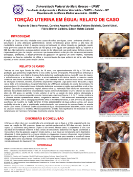
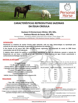
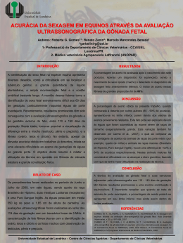
AVALIAÇÃO DA JUNÇÃO ÚTERO PLACENTA EM ÉGUAS PURO
SANGUE INGLÊS EM UM CRIATÓRIO NA REGIÃO DE BAGÉ-RS
FINGER, Ilusca1; SANTOS, Rodrigo1; LINS, Luciana2; NOGUEIRA, Carlos Eduardo3
1Acadêmico
(a) em Medicina Veterinária - FV/UFPel;2 Mestranda em Medicina Veterinária - FV/UFPel; 3 Prof. Dr.
Departamento de Clínicas Veterinária - FV/UFPel.Campus Universitário s/n°- Caixa Postal 354 - CEP 96010-900.
[email protected]
INTRODUÇÃO
RESULTADOS E DISCUSSÃO
No presente estudo uma égua (0,6%), aos 300 dias de gestação, apresentou maior espessamento
da JUP de acordo com as médias obtidas para este plantel analisado, nascendo um potro com um
quadro de asfixia neonatal. Na avaliação microscópica da placenta observou-se infiltrado inflamatório na
estrela cervical e no cordão umbilical, e também ausência de vilosidades na estrela cervical (Figura 3).
Os valores médios obtidos para a espessura da JUP das éguas estão demonstradas na tabela
abaixo:
Ultrassonografia
Trans-retal da JUP
Pode-se identificar éguas com alteração placentária durante a gestação tardia
através da medição da junção útero-placentária. A aboradagem trans-retal
permite um diagnóstico mais confiável, já que mais de 90% das infecções
placentárias são de origem ascendente.
Valores JUP
Valores referência
(Renaudin et al.,1997)
145 dias
3,28mm
3,58mm
>160 dias
3,38mm
3,84mm
>225 dias
4,72mm
>8mm
Dias gestação
OBJETIVO
O objetivo deste trabalho foi avaliar a espessura da JUP de éguas Puro Sangue Inglês (PSI)
em um criatório em Bagé-RS.
MATERIAIS E MÉTODOS
168 éguas PSI
Ultrassonografia
trans-retal
Medição da JUP
Figura
3-a.
Imagem
demonstrando
vilosidades coriônicas íntegras (entre setas).
145 dias
gestação
185 dias
gestação
215 dias
gestação
Figura 2. Superfície de uma placenta com
extensas
áreas
de
congestão
caracterizando placentite.
Figura 3-b. Imagem demonstrando
necrose das vilosidades coriônicas (entre
setas).
CONCLUSÕES
O acompanhamento da gestação em éguas a partir do quinto mês de gestação é valido
para obter-se um diagnóstico precoce de alterações placentárias.
REFERÊNCIAS BIBLIOGRÁFICAS
Figura 1. Medição da junção útero-placenta por abordagem trans-retal.
A medição é feita entre o espaço alantóide (anecóico) e o ramo médio
da artéria uterina (seta).
http://www.ufpel.edu.br/fvet/clineq
COLON, J.L. Trans-rectal ultrasonographic appearance of abnormal combined utero-placental thickness in lateterm gestation and its incidence during routine survey in a population of thoroughbred mares (2005–2008). In:
Proceedings of the 54th Annual Convention of the American Association of Equine Practitioners, San
Diego, 2008, v 54:279-285.
LE BLANC, M. M.; MACPHERSON, M.; and SHEERIN, P. (2004) Ascending placentitis: what we know about
pathophysiology, diagnosis, and treatment. In: Proceedings of the 50th Annual Convention of the American
Association of Equine Practitioners, Denver, 2004, v.50, 127-143.
MCKINNON, A. O. (2009) Maintenance of pregnancy.In: Proceedings of the 11th Annual Resort Symposium of
the American Association of Equine Practitioners AAEP, Gold Coast, 2009,v.11, 81-117.
RICKETTS, S. Management of the infertile/ subfertile mare. In: Proceedings of the
RENAUDIN,C.D.; TROEDSSON, M.H.T.; GILLIS,C.L.; KING,V .L .; and BODENA, A. Ultrasonographic Evaluation
of The Equine Placenta By Transrectal and Transabdominal Approach In The Normal Pregnant Mare. IN:
Therigenology.1997, v.47, 559-573.
10th International Congress of World Equine Veterinary Association, Moscow, 2008, v.10, 244-256.
SERTICH P.L. Clinical anatomy and evaluation of equine fetal membranes. In: Proceedings. Annu Meeting Soc
Theriogenology, 1993, v.46, 178-184.
TROEDSSON, M.; SAGE, A.M. Fetal/Placental evaluation in the mare. In: Recent Advances in Equine
Reproduction, B. Ball (Ed). Publisher: International Veterinary Information Service, Ithaca, New York, 2001.
TROEDSSON, M.H.T.; RENAUDIN, C.D.; ZENT, W.W.; STEINER, J.V. Transrectal ultrasonography of the placenta
in normal mares and mares with pending abortion: A fiel study. In: Proceedings of the 43th Annual Resort
Symposium of the American Association of Equine Practitioners AAEP, 1997, v.43, 256-258.
Download