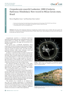
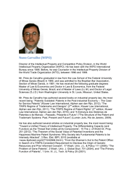
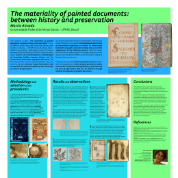
Rev. Inst. Med. Trop. Sao Paulo
53(1):39-44, January-February, 2011
doi: 10.1590/S0036-46652011000100007
ANTEMORTEM DIAGNOSIS OF HUMAN RABIES IN A VETERINARIAN INFECTED WHEN HANDLING A
HERBIVORE IN MINAS GERAIS, BRAZIL
Mariana Gontijo de BRITO(1), Talita Leal CHAMONE(1), Fernando José da SILVA(2), Marcelo Yohito WADA(3), Alexandre Braga de MIRANDA(4),
Juliana Galera CASTILHO(5), Maria Luiza CARRIERI(5), Ivanete KOTAIT(5) & Francisco Leopoldo LEMOS (1)
SUMMARY
The Ministry of Health’s National Human Rabies Control Program advocates pre-exposure prophylaxis (PEP) for professionals
involved with animals that are at risk of contracting rabies. We report an antemortem and postmortem diagnosis of rabies in a
veterinarian who became infected when handling herbivores with rabies. The antemortem diagnosis was carried out with a saliva
sample and a biopsy of hair follicles using molecular biology techniques, while the postmortem diagnosis used a brain sample and
conventional techniques. The veterinarian had collected samples to diagnose rabies in suspect herbivores (bovines and caprines) that
were subsequently confirmed to be positive in laboratory tests. After onset of classic rabies symptoms, saliva and hair follicles were
collected and used for antemortem diagnostic tests and found to be positive by RT-PCR. Genetic sequencing showed that the infection
was caused by variant 3 (Desmodus rotundus), a finding confirmed by tests on the brain sample. It is essential that professionals
who are at risk of infection by the rabies virus undergo pre-exposure prophylaxis. This study also confirms that molecular biology
techniques were used successfully for antemortem diagnosis and therefore not only allow therapeutic methods to be developed, but
also enable the source of infection in human rabies cases to be identified accurately and quickly.
KEYWORDS: Human rabies; Non-bite rabies exposure; Antemortem and postmortem diagnosis; Molecular biology.
INTRODUCTION
Rabies is an acute, progressive, fatal disease caused by a neurotropic
RNA virus from the family Rhabdoviridae, genus Lyssavirus. Mammals
from the orders Carnivora and Chiroptera are the main reservoirs13.
According to WHO estimates, around 55,000 people throughout the
world die from the disease every year, 99% of whom are on the Asian
and African continents16.
In Brazil, epidemiological investigations and laboratory diagnosis are
essential to guide measures implemented as part of the National Human
Rabies Control Program.
In Minas Gerais as well as Brazil as a whole, the incidence of human
rabies transmitted by cats and dogs has been falling over the years.
However, there has been a change in the epidemiological profile of the
disease, and cases of human rabies transmitted by bats have been reported.
dogs also fell, and the last case was reported in 2003. In 2004 and 2005,
one case of human rabies was reported each year; epidemiological and
laboratory investigation of both cases confirmed the disease had been
transmitted by hematophagous bats.
The progressive increase in the incidence of bovine rabies in Minas
Gerais is evidence of the change in the epidemiological profile of rabies,
which is reflected in the increasing importance of transmission by
hematophagous bats and other wild animals1,2,14.
We describe a case of human rabies in 2006 in the municipality
of São João del Rei, Minas Gerais, involving a health professional (a
veterinarian) who had not previously been immunized against rabies and
who refused a post-exposure regimen after he came into contact with a
herbivore that subsequently died and was confirmed in laboratory tests
to have been infected with the rabies virus.
MATERIALS AND METHODS
Although there was an increase in the number of samples sent for
laboratory diagnosis of canine and feline rabies in Minas Gerais from
2000 to 2006, the number of confirmed cases of rabies in these species fell
during the same period. The number of human rabies cases transmitted by
Epidemiological investigation and clinical data: The data were
obtained from the National Information System for Notifiable Diseases
(SINAN) and the patient’s medical records. The epidemiological
(1) SES Health Surveillance Department/MG, Belo Horizonte, Av. Afonso Pena 2300, B. Funcionários, 30130-007 Belo Horizonte, MG, Brazil.
(2) São João Del Rei Regional Department of Health. Av. Amilcar Savassi S/N, Sericícola, 36200-494 São João Del Rei, MG, Brazil.
(3) Department of Health Surveillance/MS, Brasília/DF, SCS, quadra 04, bloco A, lote 67/97, 70304-000 Brasília/DF, Brazil.
(4) Eduardo de Menezes Hospital, Rua Dr. Cristiano Resende 2213B, Bom Sucesso, 30620-740 Belo Horizonte, MG, Brazil.
(5) The Pasteur Institute of São Paulo, Av. Paulista 393, Cerqueira Cesar, 01311-000 São Paulo, SP, Brazil.
Correspondence to: Ivanete Kotait, Instituto Pasteur, Av. Paulista 393, 01311-000 São Paulo, SP, Brasil. E-mail: [email protected]
BRITO, M.G.; CHAMONE, T.L.; SILVA, F.J.; WADA, M.Y.; MIRANDA, A.B.; CASTILHO, J.G.; CARRIERI, M.L.; KOTAIT, I. & LEMOS, F.L. - Antemortem diagnosis of human rabies
in a veterinarian infected when handling a herbivore in Minas Gerais, Brazil. Rev. Inst. Med. Trop. São Paulo, 53(1): 39-44, 2011.
investigation, which was carried out by a staff from the local Department
of Agriculture, involved the patient’s family and neighborhood.
On May 15, the Minas Gerais Department of Health was notified
of a suspected rabies case in a patient admitted to Eduardo de Menezes
Hospital, a reference hospital in the state for infectious and contagious
diseases. The patient was a 27-year-old male from Campo das Vertentes,
state of Minas Gerais, Brazil.
Onset of the disease occurred on May 7, 2006, with occipital
headache and pain that radiated to the right side and was predominant in
the upper limb, the area where the patient had probably come into contact
with material from the infected herbivore. The signs evolved to include
mental confusion and a reduced level of consciousness.
The patient was put in deep sedation induced with ketamine and
midazolam, and antiviral drugs were administered (ribavirin and
amantadine). After developing intense polyuria, hyponatremia and
episodes of cardiac arrhythmia that were controlled with medication,
he died on May 26, 2006, as a result of reentrant arrhythmias that were
difficult to control, followed by cardiac arrest (Fig. 1).
Laboratory diagnosis by RT-PCR and DNA sequencing
RT-PCR: An approximately 1 cm2 sample of neck-skin biopsy and
a saliva sample were collected from the patient before his death, and a
brain sample was collected postmortem from the same patient.
Total RNA was extracted from the neck-skin sample (cut into small
pieces with a scalpel), from the brain sample and from 300 µL of saliva,
using TRIzol® reagent (Invitrogen) according to the manufacturer’s
instructions. Aliquots of a “Challenge Virus Standard” (CVS) strain
of fixed virus and water were used as positive and negative controls,
respectively. RT-PCR was carried out with sense primer 504 (5’- TATACT
CGAATCATGATGAATGGAGGTCGACT -3’) and antisense primer 304
(5’-TTGACGAAGATCTTGCTCAT-3’) for antemortem diagnosis and
sense primer 21G (5’-ATGTAACACCTCTACAATG-3’) and antisense
primer 304 (TTGACGAAGATCTTGCTCAT) for postmortem diagnosis
as previously described by MACEDO et al. (2006)10.
The RT-PCR products (249 bp and 1478 bp for antemortem and
Fig. 1 - Timeline of the course of rabies in the patient, Minas Gerais, Brazil, 2009.
40
postmortem diagnosis, respectively) were visualized under UV light
after gel electrophoresis on 1% agarose gel containing ethidium bromide
in TBE buffer.
DNA sequencing: The amplified DNA fragments were purified with
the GFXTM PCR DNA and Gel Band Purification kit (GE Healthcare),
visually quantified with a Low DNA Mass Ladder (Invitrogen) and
sequenced using the BigDye® Terminator v3.1 Cycle Sequencing Kit
(Applied Biosystems) with the sense and antisense primers according to
the manufacturer’s instructions. The reaction products were then resolved
in an ABI-3130 automatic sequencer (Applied BiosystemsTM).
Phylogenetic analysis: For the antemortem diagnosis, a 249 bp
region of the nucleoprotein (N) gene located between nucleotides
1286 and 1533 of the Pasteur Virus (PV) (GenBank accession number
M13215.1) was analyzed, while for the postmortem diagnosis a 1478
bp region of the N gene located between nucleotides 55 and 1533 of the
PV virus was analyzed. First, the raw sequencing data were edited using
CHROMAS version 2.24 software (Copyright© 1998-2004 Technelysium
Pty Ltd.), and the final consensus sequence for each sample (neck-skin,
saliva and brain tissue) was aligned with homologous sequences in
GenBank using the CLUSTAL/W method with the Bioedit program and
submitted to BLASTn to confirm sequence identity7. The alignment was
then used to build a neighbor-joining distance-based phylogenetic tree
using the Kimura two-parameter correction model with 1,000 bootstrap
replicates and the Mega 2.1 program8. The identities between the aligned
sequences were calculated using the Bioedit program.
Direct immunofluorescence: This was carried out using a
fluorescein-conjugated anti-rabies polyclonal antibody produced in
hyperimmunized rabbits by the Pasteur Institute of São Paulo and slides
prepared with impressions of brain tissue, as described by DEAN et al.
(1996).
Viral isolation in mice: A suspension was prepared from the brain
sample and inoculated intracerebrally in eight 21-day-old Swiss albino
mice, according to the technique advocated by KOPROWSKI (1996)9
Antigenic typing by indirect immunofluorescence using
monoclonal antibodies: Slides prepared with brain tissue from firstpassage mice were typed antigenically using a panel of monoclonal
BRITO, M.G.; CHAMONE, T.L.; SILVA, F.J.; WADA, M.Y.; MIRANDA, A.B.; CASTILHO, J.G.; CARRIERI, M.L.; KOTAIT, I. & LEMOS, F.L. - Antemortem diagnosis of human rabies
in a veterinarian infected when handling a herbivore in Minas Gerais, Brazil. Rev. Inst. Med. Trop. São Paulo, 53(1): 39-44, 2011.
antibodies provided by the Centers for Disease Control and Prevention
(CDC), in Atlanta, USA, to characterize the variants isolated on the
American continent6. Serological testing could not be carried out because
serum samples were not collected at any stage of the disease.
RESULTS
Epidemiological investigation and clinical data: Exposure probably
occurred in March 2006 in the town of Prados, state of Minas Gerais,
Brazil, and it was suggested that the virus might have been transmitted
by an animal from the caprine species that the patient, a veterinarian,
was attending to. The municipality where he had been working had had
several cases of rabies in herbivores, all confirmed by laboratory tests
at the Minas Gerais Institute of Agriculture (IMA). It was not possible,
however, to identify the species of herbivore involved in the infection
or the exact date and time that the infection occurred. The veterinarian
worked on various farms in the municipalities of Prados, Coronel Xavier
Chaves, Tiradentes and São João del Rei. In the same month there were
four cases of rabies in herbivores (three in bovines and one in a caprine)
in the municipality of Prados. There were also two confirmed cases in
bovines in other municipalities in the São João del Rei Regional Health
Department district; one of these was in Madre de Deus de Minas, and
the other in Entre Rios de Minas. According to information provided by
the IMA (personal information), there had been clinical cases of rabies
in herbivores in various municipalities in the region, although these had
been neither reported nor confirmed by laboratory tests.
The patient did not report having been attacked by bats, dogs or other
animals and had never had either pre-exposure antirabies vaccinations or
antirabies prophylaxis after contact with suspect herbivores. He did not
use any type of personal protection equipment to collect samples from
animals suspected of rabies.
Laboratory diagnosis
RT-PCR, DNA sequencing and phylogenetic analysis: The neckskin biopsy and saliva samples were positive by RT-PCR, yielding a 249
bp fragment, and the brain sample, which was also positive by RT-PCR,
yielded a 1478 bp fragment. No amplified products were detected in the
negative control, and no extra band was detected. A total of 249 and 1478
nucleotides located between nucleotides 1286/1533 and 55/1533 of the
rabies virus genome, respectively, were analyzed using the PV strain as
a reference. Final sequences of 106 and 1290 nucleotides of the N-gene
region between nucleotides 1314-1420 and 130-1420 of the PV virus
were obtained for antemortem and postmortem diagnosis, respectively.
The topology of the phylogenetic tree (106 bp - antemortem
diagnosis) showed four distinct clusters (Fig. 2): 1 - samples associated
Fig. 2 - Phylogenetic tree based on the sequence of 106 nucleotides of the rabies virus N gene between positions 1314-1420. Phylogenetic analysis was performed using the neighbor-joining
method. Isolates IP3895/neck-skin biopsy and saliva and IP4435/Central Nervous System, which are shown underlined and bold, segregated into the same group related to viruses isolated in
hematophagous bats (Desmodus rotundus). The names of the samples in the tree refer to the GenBank accession numbers.
41
BRITO, M.G.; CHAMONE, T.L.; SILVA, F.J.; WADA, M.Y.; MIRANDA, A.B.; CASTILHO, J.G.; CARRIERI, M.L.; KOTAIT, I. & LEMOS, F.L. - Antemortem diagnosis of human rabies
in a veterinarian infected when handling a herbivore in Minas Gerais, Brazil. Rev. Inst. Med. Trop. São Paulo, 53(1): 39-44, 2011.
Fig. 3 - Phylogenetic tree based on the sequence of 1290 nucleotides of the rabies virus N gene between positions 130-1420. Phylogenetic analysis was performed using the neighbor-joining
method. Isolates IP4435/Central Nervous System, which are shown underlined and bold, segregated into the same group related to viruses isolated in hematophagous bats (Desmodus rotundus).
The names of the samples in the tree refer to the GenBank accession numbers.
with hematophagous bats; 2 - samples associated with insectivorous bats;
3 - fixed samples; and 4 - samples associated with dogs.
Isolates IP3895 (neck-skin biopsy and saliva) and IP4435 (brain tissue)
had 100% identity between them and segregated into a cluster formed by
Desmodus rotundus isolates (bootstrap = 97) from different regions of Brazil.
The topology of the phylogenetic tree (1290 bp - postmortem
diagnosis) showed four distinct clusters (Fig. 3): 1 - associated with
hematophagous bats; 2 - samples associated with insectivorous bats;
3 - fixed samples; and 4 - samples associated with dogs.
Isolate IP4435 (brain tissue) segregated into a cluster formed by
Desmodus rotundus isolates (bootstrap = 100) from different regions
of Brazil.
Direct immunofluorescence: The slides were positive for the rabies
antigen and the test was specific as no fluorescence was detected in the
negative control.
Viral isolation in mice: The virus was isolated 10 days after
inoculation of the mice.
Antigenic typing by indirect immunofluorescence using
monoclonal antibodies: The reactivity pattern of the virus isolated from
mice was compatible with that of variant 3 (AgV3), which is characteristic
of the variant isolated from the hematophagous bat Desmodus rotundus,
the main transmitter of rabies to herbivores.
42
DISCUSSION
With the increasing number of rabies cases in economically important
animals, veterinarians or animal owners who handle these animals are
at risk of contracting the disease since, irrespective of the species they
belong to, infected animals eliminate the virus in their saliva during the
pre-symptomatic and symptomatic phases3,5.
The Brazilian Ministry of Health Regulations for the Prophylaxis of
Human Rabies recommend that individuals whose work exposes them
to a risk of contracting rabies through contact with animals receive
pre-exposure prophylaxis (PEP) and that those who have been exposed
to animals with rabies receive post-exposure vaccination. In the case
described here, the patient had not received PEP and had refused postexposure prophylaxis; this lack of immunological response led to his
death11.
Exposure probably occurred in March 2006 in the town of
Prados, Minas Gerais, and the likely transmitter was a caprine that
the veterinarian had been attending to. He had been working in the
municipality of Prados, where there had been cases of animal rabies that
had been confirmed in laboratory tests. However, the type of herbivore
and exact place and time the infection occurred could not be determined
with any degree of certainty as the patient had been working on various
farms in the region.
As there was no record of the veterinarian having been bitten by
bovines or caprines, exposure must have occurred when he was handling
BRITO, M.G.; CHAMONE, T.L.; SILVA, F.J.; WADA, M.Y.; MIRANDA, A.B.; CASTILHO, J.G.; CARRIERI, M.L.; KOTAIT, I. & LEMOS, F.L. - Antemortem diagnosis of human rabies
in a veterinarian infected when handling a herbivore in Minas Gerais, Brazil. Rev. Inst. Med. Trop. São Paulo, 53(1): 39-44, 2011.
infected animals and came into contact with their saliva. The records
show that liquids were administered orally to an infected animal in
March. Contamination may also have occurred when a brain sample was
being collected to be sent to the diagnostic laboratory for confirmation
of suspected rabies. On both occasions the veterinarian failed to use
personal protection equipment (PPE).
The present study describes the first human rabies case successfully
diagnosed antemortem using molecular biology methods standardized
by MACEDO et al. (2006)10. The recent cases of rabies in Wisconsin,
USA, and Recife, Brazil, in which patients survived when coma was
induced and antiviral drugs were used, show that molecular methods are
important tools for the early identification of human rabies and allow the
Milwaukee15 or Recife12 protocols to be used.
Antigenic and genetic studies identified variant 3 - compatible with
the variant found in the hematophagous bat Desmodus rotundus, the
main transmitter of rabies in herbivores - in the saliva, hair follicles and
brain tissue of the patient, confirming the data from the epidemiological
investigation.
Efforts should be made by federal, state and municipal health and
agricultural institutions as well as by universities to make people aware
that the antirabies vaccine is strongly recommended for veterinarians
and can help avoid cases like that described here.
We conclude that:
1. In our study the molecular biology methods currently available
for antemortem laboratory diagnosis of rabies were used successfully.
These allow therapeutic methods for the treatment of human rabies
to be developed and maintained and the source of infection in rabies
cases to be accurately identified before death, so that a more thorough
epidemiological investigation can be undertaken.
2. It is essential that professionals who are at risk of infection by the
rabies virus undergo PEP to ensure they are appropriately immunized for
field activities such as the collection of samples to diagnose animal rabies.
3. Individuals exposed to suspect animals, particularly those that
have been confirmed rabies-positive in laboratory diagnostic tests,
must undergo post-exposure prophylaxis as advocated by the Brazilian
Ministry of Health and World Health Organization.
4. Educational programs focusing on PEP and appropriate serological
follow-up should be developed with universities and veterinary services.
RESUMO
Diagnóstico ante-mortem de raiva humana em médico veterinário
infectado por manipulação de herbívoro, Minas Gerais, Brasil
O Programa Nacional de Controle da Raiva Humana do Ministério
da Saúde preconiza o esquema profilático pré-exposição (PEP) para
profissionais envolvidos com animais expostos ao risco de contraírem
raiva. O presente trabalho relata o diagnóstico de raiva (ante e postmortem) em veterinário infectado por manipulação de herbívoros
raivosos. O diagnóstico laboratorial ante-mortem foi efetuado a partir
da saliva e biópsia de folículo piloso, utilizando técnicas de biologia
molecular e o post-mortem a partir do tecido cerebral e de técnicas
convencionais. O médico veterinário coletou amostras para diagnóstico de
raiva em herbívoros (bovinos e caprinos) suspeitos que, posteriormente,
foram confirmados positivos em laboratório. Após a apresentação dos
sintomas clássicos de raiva e realizadas as provas de diagnóstico antemortem com saliva e folículo piloso, ambas as amostras apresentaram
resultados positivos pelo nested-RT-PCR. O sequenciamento genético
revelou que a infecção se deu pela variante 3 do Desmodus rotundus,
resultados estes confirmados com a amostra do cérebro. É indispensável
que profissionais expostos ao risco de infecção pelo vírus da raiva
realizem a profilaxia pré-exposição. Ressalta-se, também, que as técnicas
de biologia molecular apresentaram bons resultados para a realização de
diagnóstico ante-mortem, propiciando o desenvolvimento de métodos
terapêuticos, e determinando com precisão e rapidez a fonte de infecção
dos casos de raiva humana.
REFERENCES
1. Barbosa AD, Silva JA, Moreira EC, Meneses JNC, Magalhães DF, Menezes FL et al.
Distribuição espacial e temporal da raiva canina e felina em Minas Gerais, 2000 a
2006. Arq Bras Med Vet Zoot. 2008;60:837-42.
2. Brito MG, Chamone TL. Ações de controle da raiva canina e felina no estado de Minas
Gerais, 1999-2002. Bol Epidemiol. 2002;6:1-8.
3. Carrieri ML, Peixoto ZM, Paciência ML, Kotait I, Germano, PM. Laboratory diagnosis
of equine rabies and its implications for human postexposure prophylaxis. J Virol
Methods. 2006;138:1-9.
4. Dean DJ, Albelseth MK, Atanasiu P. The fluorescent antibody test. In: Meslin, FX, Kaplan
MM, Koprowsky H, editors. Laboratory techniques in rabies. 4th ed. Genebra: World
Health Organization; 1996. p. 88-95.
5. Delpietro HA, Larghi OP, Russo RG. Virus isolation from saliva and salivary glands of
cattle naturally infected with paralytic rabies. Prev Vet Med. 2001;48:223-8.
6. Diaz AM, Papo S, Rodriguez A, Smith, JS. Antigenic analysis of rabies-virus isolates
from Latin American and the Caribbean. Zentralbl Veterinarmed B. 1994;41:153-60.
7. Hall TA. Bioedit: a user-friendly biological sequence alignment editor and analysis
program for Windows 95/98/nt. Nucl Acids Symp Ser. 1999;41:95-8.
8. Kumar S, Tamura K, Jakbosen IE, Nei M. Mega 2: molecular evolutionary genetic analysis
software. Tempe: Arizona State University; 2001.
9. Koprowski H. The mouse inoculation test. In: Meslin FX, Kaplan MM, Koprowsky H,
editors. Laboratory techniques in rabies. 4th ed. Genebra: World Health Organization;
1996. p. 80-7.
10. Macedo CI, Carnieli Jr P, Brandão PE, Travassos da Rosa ES, Oliveira RN, Castilho JG,
et al. Diagnosis of human rabies cases by Polymerase Chain Reaction of neck-skin
samples. Braz J Infect Dis. 2006;10:341-5.
11. Ministério da Saúde. Fundação Nacional de Saúde. Guia de vigilância epidemiológica.
5a ed. Brasília: Ministério da Saúde; 2002. 2 v.
12. Ministério da Saúde. Protocolo para tratamento de raiva humana no Brasil. Epidemiol
Serv Saúde. 2009;18:385-94.
13. Rupprecht CE, Hanlon CA, Hemachudha T. Rabies re-examined. Lancet Infect Dis.
2002;6:327-43.
14. Schneider MC, Belloto A, Adé MP, Hendrickx S, Leanes LF, Rodrigues MJF, et al. Current
status of human rabies transmitted by dogs in Latin America. Cad Saúde Pública.
2007;23:2049-63.
43
BRITO, M.G.; CHAMONE, T.L.; SILVA, F.J.; WADA, M.Y.; MIRANDA, A.B.; CASTILHO, J.G.; CARRIERI, M.L.; KOTAIT, I. & LEMOS, F.L. - Antemortem diagnosis of human rabies
in a veterinarian infected when handling a herbivore in Minas Gerais, Brazil. Rev. Inst. Med. Trop. São Paulo, 53(1): 39-44, 2011.
15. Willoughby RE, Tieves KS, Hoffman GM, Ghanayem NS, Amlie-Lefond CM, Schwabe
MJ, et al. Survival after treatment of rabies with induction of coma. N Engl J Med.
2005;352:2508-14.
16. World Health Organization. WHO Expert Consultation on Rabies. First report. Geneva:
World Health Organization; 2005. 121p. (Technical Report Series; 931).
Received: 22 March 2010
Accepted: 22 November 2010
44
Download